KOMPOZİT DOLGU

Kompozit diş dolgularının dişi çürütebildiği, sinirini öldürebildiği kanser yapabildiği, gebelik ve emzirmede sakıncalı olduğu, kısa ömürlü olduğu, ağızdaki mikropları besleyebildiği anlatıldı.
Özet:
Diş dolgusu yapılırken dişe sürülen bond ve dolgu olarak kullanılan resin esaslı adezivlerin yapısında metakrilat monomerleri bulunur. Bunlar genel olarak :
• Sitotoksik, genotoksiktir, ve serbest oksijen radikali oluşturur,
• DNA hasarı yapar, teratojendir,
• Bakteriler tarafından beslenme kaynağı olarak kullanılır, bakterileri çoğaltır,
• Pulpada tahriş ve nekroz oluşturur (kompozit nekrozu),
• Pulpanın iyileşmesini geciktirir,
• Çürük yapıcı mikropların üremesine yardımcı olur,
• Temas ettiği hücrenin intihar etmesine yol açar,
• Amalgam dolgudan daha kısa ömürlüdür,
• Sertleşirken büzülür, kavite kenarında sızıntıya yol açan mikro boşluklar oluşturur,
• Salyada çözünür, eksilir, zaman içinde giderek yumuşar ve dağılır,
• Tıkanma tam gerçekleşmez, daima dolgunun kenarında sızıntı yapar,
• Obesiteye sebep olabildiği söylenmektedir,
• Diyabete eğilim yaratabilir,
• Kadınlık hormonu (östrojen) etkisi gösterir, üreme (rahim prostat, meme), akciğer ve beyin kanserleri ile ilişkilidir,
• Salyadaki nötrofiller kompozit dolguya saldırır ve dolguyu tahrip eder.
• Kompozit dolgu sökülürken hekim ve sağlık personeli özel önlem alarak kendisini korumalıdır.
Amaç
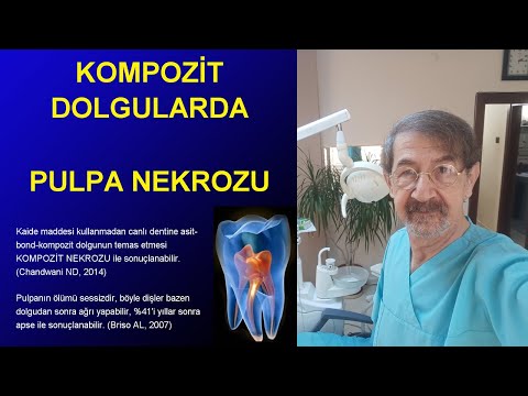
Resin esaslı adeziv restorasyonlar, pulpa ve vital dentinin iyice korunmadığı veya ışıkla polimerizasyonun yetersiz yapıldığı durumlarda belirtisiz pulpa ölümlerine sebep olabilir. Asemptomatik dönemde hasta ve hekimin bu hasarın farkına varması zordur. Hemen gelişen aseptik pulpa nekrozu ile akut infeksiyonun başlaması arasında yıllarla ölçülebilen uzun bir sükunet dönemi bulunabilir. Klinik şikayetlerin geç başlaması sebebi ile resin restorasyonlu bir dişin apsesinin kompozit nekrozu sebebi ile oluştuğunu ayırt etmek her zaman mümkün olmayabilir. Neden nekroz geliştiği, nekrozun tedavisi üzerine belirleyici de değildir. Adeziv restorasyonlu dişlerde hemen başlayan postop ağrı veya herhangi bir zamanda başlayan akut infeksiyonlara, kompozit nekrozunun sebep olabileceği daima hatırlanmalıdır.
Adezif dolgular yapılırken etch asit, bond ve dolgu materyalinin vital dentine temas etmesinden kaçınmak veya pulpayı korumak gerekir. Işıkla polimerizasyon süresinin önerilenden daha uzun olması uygun bir tedbir olabilir. Posteryor restorasyonlarda, yüksek çürük ve sızıntı riski olan ve fazla basınç alacağı bilinen dişlerde kompozit dolgu yerine, amalgam dolgu daha başarılı bulunmuştur.
Bu çalışmada kompozit dolguların pulpa nekrozundan, yumuşak doku tahrişine kadar geniş bir skalada lokal selüler zararları ve subklinik seyrediyor olabilecek sistemik toksisitelerine mercek tutuldu. Bu malzemelerin zamanla yumuşadıkları, bakterilerin çoğalmasını destekledikleri, sekonder çürük geliştirdikleri, kanserojen, mutajen, teratojen etkileri ele alınmıştır. Amalgam dolguya alternatif olarak ortaya çıkmış olmalarına rağmen oldukça eksiktir. Amalgam dolgu yerine kompozit dolgular bir alternatif olmaktan uzak görünmektedir veya kompozit dolgulara bir alternatif bulunmasının gerektiği açıktır. Azılar bölgesinde kompozit dolguya en iyi alternatif amalgam dolgu olarak görünmektedir.
Bu makalenin amacı:
★ Kompozit dolguların pulpa nekrozu yapabildiğine dikkat çekmek, kaide maddesinin mutlaka kullanılması gerektiğini hatırlatmaktır.
★ Başta gebeler, süt verenler ve çocuklar olmak üzere her bireye kompozit dolgu yapmanın riskli olabileceğini vurgulamaktır Başka materyali tercih etmenin daha makul olabileceğini hatırlatmaktır.
Bu konuda literatür bulguları aşağıda tartışılmıştır.
Giriş
Adeziv resin sistem dental restorasyon materyalleri giderek artan sıklıkta ve çeşitlilikte dünyada yaygın olarak kullanılmaktadır. Bütün adeziv sistemlerin yapısında genellikle aynı kimyasallar bulunur. BisGMA ve türevleri olan bir düzine civarında resin esaslı madde her marka kompozit dolguların ana bileşenidir. Bu gün mükemmele yakın estetik doğası sebebi ile kompozit dolgular pazarda önemli bir yer tutmaktadır. Amalgam dolgulardan daha fazla yapılmakta ve satılmaktadır.
Kompozit dolguların yapısına bulunan neredeyse bütün maddeler toksik ve sağlığa aykırı olmasına rağmen yaygın şekilde dillendirilmemekte, yan tesirleri prospektüslerde yer almamakta ve toksik etkileri firmalar tarafından geri planda tutulmaktadır. (Dursun E, 2016) Hatta tam tersine bilgiler verilerek yapısında toksik madde bulunmadığı firmalar tarafından beyan edilmektedir. Oysaki yapısında toksik madde bulunmadığı iddia edilen kompozit dolgularlardan emisyonu en az 1 yıl boyunca devam eden toksik maddeler yayıldığı yapılan çalışmalarda (Roberson TM, 2006) (De Nys S, 2021) açıkça gösterilmiştir.
Bu çalışmada kompozit dolguların pulpa nekrozundan, yumuşak doku tahrişine kadar geniş bir skalada lokal, sistemik, selüler zararlarına, bakterilerin çoğalmasını destekleyen etkilerine, sekonder çürük oluşturmalarına, teratojen, mutajen ve kanserojen etkilerine, ve subklinik seyrediyor olabilecek zararlarına mercek tutuldu.
Resin esaslı adezivlerin kimyasal yapısı:
Bütün resin esaslı adezivler (kompozit dolgu, bond, fissür örtücü, kompomer, resin modifiye cam iyonomer ve diğerleri) değişik kombinasyonlarda olarak şu maddelerden imal edilmektedir.
Bond ve kompozit dolgunun yapısında bulunan maddeler (alfabetik):
10-MDP, 10-methacryloyloxydecyl dihydrogen phosphate,
4-META, 4-methacryloxyethyl trimellitate anhydride,
4-MET, 4-methacryloxyethyl trimellitic acid,
BADGE, Bisphenol A diglycidyl ether,
BHT, 2,6-di-tert-butyl-p-cresol,
BisDMA, Bisphenol A dimethacrylate,
Bis-EMA, ethoxylated BisGMA,
Bis-GMA, bisphenol A–glycidyl methacrylate,
BPA, Bisphenol A,
CQ, Camphorquinone,
CEMA, N-(2-cyanoethyl)-N-methylaniline ,
DEGDMA, diethylene glycol dimethacrylate,
DMABEE, 4-N,N-dimethyl amino benzoic acid ethylester,
DMAEMA, 2-(dimethylamino)ethyl methacrylate,
DMPA, 2,2 dimethoxy-2-phenyl-acetophenone,
DPICI, diphenyliodoniumchloride DPO, dibenzoyl peroxide,
DUDMA, diurethane dimethacrylate,
EGDMA, ethylene glycol dimethacrylate,
EMPA, 2,3-epoxy-2-methylpropionic acid,
EMPME, 2,3-epoxy-2-methyl-propionicacid-methylester ,
GDMA, glycerol-dimethacrylate,
GPDMA, glycerol phosphate dimethacrylate,
HEMA, 2-hydroxyethyl methacrylate,
HMBP, 2-hydroxy-4-methoxy-benzophenone,
MA, methacrylic acid,
MDTP, 10-methacryloyloxydecyl dihydrogen thiophosphate,
MPC, 2-methacryloyloxyethyl phosphorylcholine ,
MTU-6, 6-methacryloyloxyhexyl 2-thiouracil 5-carboxylate,
PENTA, dipentaerythritol penta-acrylate phosphate,
TEGDMA, triethylene glycol dimethacrylate,
TPO, diphenyl (2,4,6-trimethylbenzoyl) phosphinoxide,
UDMA, urethane dimethacrylate.
Kompozit dolgunun yapısı
Kompozit dolgular prensip olarak üç tür bileşenden imal edilir:
1) polimerize olabilen organik resin monomerler, örneğin Bis-GMA, UGDMA, TEGDMA veya EGDMA.
2) Boşluk doldurucu inorganik maddeler ve
3) Organik ile inorganik bileşenler arasında birleşmeyi sağlayan silan moleküllerinden oluşur. Poliasitler ile modifiye edildiğinde bunlara "kompomer" adı verilir.
 A- 2,2-bis[4(2-hydroxy-3-methacryloxypropoxy)-phenyl]propane (BisGMA);
B- n=1, Ethylene glycol dimethacrylate (EGDMA); n=2, Dithylene glycol dimethacrylate (DEGDMA); n=3,Triethylene glycol dimethacrylate (TEGDMA);
C- Urethane dimethacrylate (UDMA);
D- Ethoxylated bisphenol A based dimethacrylate (Bis-EMA)
Her marka bütün kompozit dolgular bu maddeden veya bunun türevlerinden imal edilir. Bunlar aslında bir monomerdir. Monomerlerin uc-uca birleşmesi foto initiyatör maddeler ile gerçekleşmektedir. Uçları birleştiğinde zincirleşir, polimerleşir.( Delaviz Y, 2013)
Bondun pulpaya zarar vermesi
Bondların yapısı kompozit dolgudan farklı değildir. Çünkü "bond, aslında sıvı kompozit"tir. Piyasamızdaki bondların yapısı her markaya göre küçük değişiklikler göstermekle birlikte esas olarak methacrylate, fosforize edilmiş pentacrylate, aldehit gibi toksik ve kanserojen ethylene glikol, EGDMA, DEGDMA ve 10-MDP türevlerinden oluşur ve pH derecesi 1-3 arasındadır kuvvetli bir asittir. (Cadenaro M, 2023) (Tuncer S, 2012) En az asit olanı bile pH 3 seviyesinde güçlü bir asittir. Böyle kuvvetli bir asitin pulpayı tahriş etmemesi düşünülemez. Aşağıdaki tabloda piyasamızda bulunan bondların (en fazla asit olandan en az asit olana doğru sıralanmış olarak) içerikleri görülmektedir. Bondların hepsinin kuvvetli bir asit olduğuna (pH sütununa) ve çözücü olarak alkol aseton içerdiklerine dikkat ediniz.

Adezivlerin asitlik dereceleri
(Tıklanırsa resim büyür)
Bu tabloda listelenen resin bazlı kompozit materyallerin hepsi fevklade toksiktir. Bis-GMA isimli madde, HEMA ve TEGDMA isimli maddelerden 133 kat daha toksiktir (Yıldız M, 2017) Kompozitlerde genel kural olarak moleküler ağırlığı daha yüksek olan daha toksiktir.
300, 500, ve 1000μ lik denin diskleri hazırlanmış, dentinin oklüzal tarafına şu maddeler 24 saat boyunca temas ettirilmiştir. 1) Vitrebond markalı cam iyonomer siman, 2) GLUMA Bond5 markalı total-etching adhesive 3) GLUMA Self Etch adhesive 4) Single Bond Universal markalı adeziv. Bir günün sonunda dentin disklerinin altında kalan hücre kültüründeki canlı hücre sayıları yoklanmıştır. Vitrebond, 300 um diskin altındaki canlı hücre sayısını %10'a düşürmüştür. 500 um diskin altında canlı hücre sayısı %17,1000 um de %18 bulunmuştur. Bu dört adeziv içerisinde en sitotoksik bulunmuştur. Sonraki sırada GLUMA Bond5 gelmektedir, diğer ikisi hücre sayısını anlamlı şekilde azaltmamıştır. Bu maddelerin canlı dentine temas ettirilmemesi, araya koruyucu bir materyal kullanılmasının gerekli olduğu vurgulanmıştır. (Jiang RD, 2017)
Buna benzer sayısız deney literatürde bulunmaktadır. hepsinde adeziv materyalin şiddetli toksik doğası şüphe götürmez şekilde ifade edilmiştir.
Bir başka deneyde 500 μm kalınlığında hazırlanan dentin diskleri pulpa hücre kültürünün üzerine yerleştirilmiş ve dentin diskinin üzerine sırası ile aşağıdaki şu bondlar, 24 saat süre ile temas ettirilmiştir. Sonra pulpa hücrelerinin metabolizmaları izlenmiştir. 1- G-Bond [GB], 2- Adper Prompt Self-Etch [APSE], 3- Clearfil DC Bond System [CDCB], 4- Quadrant University-1-Bond [UB]) Bir günün sonunda pulpa hücrelerinin metabolizmalarının bozulma yönünde etkilendiği görülmüştür. Dentinin dolgudan önce korunması gerektiği ifade edilmiştir. (Sengün A, 2011)
Etch asitin pulpaya zarar vermesi:
Etch kelimesi bir kısaltma değildir. Lisani anlamı "ısırmaya veya beslenmeyi sağlayan " dır. Bir koroziv yüzey yapmak için bir asit veya başka bir aşındırıcı madde ile bir yüzeyi kesmek anlamına gelir. Güncelde, elektronik devre baskılı devre kartları ve plaketleri yapma tekniği olarak bilinir. Ancak aynı zamanda metal üzerine dekorasyon yapmak anlamı da vardır. Dolgudan önce mineye asit sürüp mineyi dekalsifiye ederek pürüzlendirme işlemine etching veya pürüzlendirme denilmiştir. Bu amaç ile kullanılan %37 fosforik asite "etch asit" denir. Resin esaslı adeziv (kompozit) dolgular yapılırken pürüzlendirme işlemi için mineye asit uygulanır, 10-30 sn bekletilir. Bu sürede mine kalsiyumunu kaybedip organik kısmı açığa çıkar. Sonra asit yıkanıp kavite kurutulur. Sırasıyla bond ve kompozit dolgu uygulanır. Eğer işlem sırası böyle oluyorsa buna "total etch" ismi verilmiştir. Bu terim çok isabetli değildir ancak yerleşik terimdir.
Asitleme sırasında minenin mineral yapısının çözünmesi ile açığa çıkan ve mine yüzeyinde ortama yayılan kalsiyum iyonlarını monomerlerin uçlarına bağlayarak adezivin dişe bağlanmasını güçlendirme fikri ortaya atılmıştır. Böylece Ca-monomer bağlantılı nano hibrit tabakalar oluşması hedeflenmiştir. Bu amaç ile hazırlanmış bir adezivin kutusunda asit ile bond aynı şişenin içinde bulunur ve tek bir solüsyon şeklinde mineye uygulanır. Bu şekilde asitlemeye "self etch" (kendinden pürüzlendiren) adı verilir.
Ortodontik braketlerin yerleştirilmesi sırasında hem total etch hem de self etch metotları uygulanabilmektedir.
Etch asitin canlı dentin dokusuna temas etmesi orta derece ile çok sert derece arasında değişen pulpa tahrişi sebebidir (Pashley DH, 1992) (Bowen RL, 1992) Maymun dişine uygulanan etch asitin pulpa üzerinde iritasyona sebep olduğu ve 3üncü günde odontoblastik tabakaya zarar verdiği, bu hasar sırasında olaya hiç bakteri karışmadığı, yıkımın bakterisiz olarak meydana geldiği görülmüştür. İritasyon 30 ve 90 ıncı günlerde azalmış fakat dentin harabiyeti kalıcı olmuştur. Asit sadece mineye temas edip dentine temas etmediğinde böyle bir hasar görülmemiştir. Geç meydana çıkan hasarın bakteri istilasına sıkıca ilişkili bulunmuştur. (Fujitani M, 1992)
Dentin etch asit ile temas edince zaten önceden geçirgen olan dentin dokusu demineralize olarak daha da geçirgen bir yapı kazanmaktadır. Bu durum gelecekte pulpanın bakteri istilasını kolaylaştırmakta ve hasarın daha ağır olmasına sebep olmaktadır. (Quist, 1989) (Fujitani, 1992)
Dentin adezivleri
Dentinin kompozite tutunmasını sağlayan dentin adezivleri (liner) ve dentin hassasiyetini gideren açığa çıkmış kök yüzeylerine uygulanan adezivler etch asit uygulaması ile başlar. Daha sonra hidrofilik monomer yani bir bond ajanı (mesela aseton ve alkol içerisinde çözünmüş HEMA) uygulanır. Bunun yapısında glutaraldehit bulunur. Bu madde dentin tüpleri içerisindeki protein lifleri fikse etmek içindir. yeterince polimerize olmamış dentin adevivlerinin kütlesinden ethylene-glycol methacrylat, EGDMA ve D/TEGDMA, H-EIMA and MMA emisyonu gösterilmiştir. (Kanerva L, 1994a)
HEMA, 14-META, BisGMA ve UDMA makrofajlatın mitokondriyal aktiviteleri üzerine denenmiştir. UDMA>Bis-GMA>4-META>HEMA şeklinde toksisite sırasına sokulmuştur. (Rakich, 1998)
Fissür koruyucu
Fissür sealent veya fissür koruyucu ismi ile bilinen materyaller aslında sıvı kompozittir. Yapısında flor salınımı yapması amacı ile flor tuzları ilave edilmiş olabilir. Ancak resin adezivlerin bütün toksik etkilerini taşır.
Radyoaktif izotopla işaretlenmiş 2 tane fissür koruyucu, 15 kişinin dişlerine uygulanmıştır. Fissür koruyucudan ağıza ve vücuda yayılan BPA'nın salya ve idrardaki konsantrasyonları ölçülmüştür. Delton LC markasında Salyada BPA 0.34 ng/ml'den 42.8'e çıkmış 1 saat sonra 7.86 ng/mL ye düşmüştür. İdrarda 2.41'den 20.1'e çıkmış 1 saat sonra 5.14ng/mL'e gerilemiştir. Helioseal F markasında salyada BPA 0.22'den 0.54'e çıkmış 1 saat sonra 0.21 'e düşmüştür. İdrarda 2.12'den 7.26'ya çıkmış 1 saat sonra 2.06 ng/mL'ye gerilemiştir. Helioseal F markanın Delton LC markadan daha az BPA yayıldığı gösterilmiştir. (Joskow R, 2006)
Fissürün kapatılması amacıyla kulanılan fissür koruyucunun yapısında bulunan BisDMA, BPA'ya dönüşmekte ve hem toksisite göstermekte hem de kendisi ile diş sert dokusu arasında sızıntı yapmaktadır. İlk 24 saat içinde BisDMA'nın BPA'ya konversiyonu hızlıdır. Sonra azalmaktadır. (Schmalz G, 1999)
Resin modifiye cam iyonomer simanlar:
İyon açığa çıkaran cam partikülleri, suda çözünebilen poliakrilik asitler, ışıkla sertleşebilen monomerler (örn. HEMA) ve katkı maddeleri içerir. Açığa çıkarması planlanan iyon genellikle florid olmuştur. Fakat kromatografi çalışmaları ortama HEMA'nın da salındığını göstermiştir. (Geurtsen, 1998b) Özellikle Vitremer markalı resin modifiye cam iyonomerden salınan HEMA nın osteoblastlar üzerine oldukça toksik olduğu gösterilmiştir. İnsan fibroblastlarında protein sentezini kalıcı olarak %100 oranında durdurabilmektedir. Bu toksik etkiyi dentin bariyerinin arkasından yapmaktadır (Olivia, 1996)
Vitrebond ve Syntaci markalı ürünlerin fotoinitiyatör madde olan DPICI saldığını bunu yeterince polimerize olmadığında chlorine, iodine, bromine ve benzen gibi oldukça toksik maddelere dekompoze olduğunu göstermişlerdir.(Geurtsen, 1999c) bu maddelerin dolgu kütlesinden dışarı sızmaları muhtemeldir. Bu konuda yeterli klinik bilgimiz yoktur.
Resin esaslı adezivlere ilave edilen fotoinitiyatör maddeler
Adezivlerin (kompozit, bond, resin) polimerize olup sertleşmesi için mavi ışığa duyarlı "fotoinitiyatör madde"ler ilave edilir. Bu maddeler mavi ışığa maruz kaldığında monomerlerin ucuca birleşerek polimerize olmasını başlatır. Bu maddeler kompozit dolgunun yapısına %0.5 w/w oranında ilave edilir. Mavi ışığa duyarlı olup reaksiyonu başlatan, bu maddelerdir. Şimdi fotoinitiyatörlere bakalım:
Fotoinitiyatör maddeler sadece resin esaslı monomerlerin değil, bond, fissür örtücü gibi diğer ışıkla reaksiyon başlatan maddelerin de yapısına ilave edilmektedir. Esas olarak 3 madde bu amaçla kullanılır.
1- Diphenyl (2,4,6 dimethyl-benzoyl) phosphine oxide (TPO) 230–430 nm de tetiklenir.
2- Camphorquinone (CQ) (C10H14O2). 400–500 nm de tetiklenir.
3- Phenyl Propanedione (PPD). 350 - 490 nm arasında tetiklenir.
Şimdi bunları bir başka sayfada
inceleyelim↗
Fotonitiyatör maddelerin canlı dokuya etkileri:
Adeziv sistemlerde kullanılan etch asit, bondu oluşturan maddeler, bondun pH derecesi, bondun çözücüsünün ayrı ayrı tahriş yaratıyor olmalarının yanında ayrıca adezivin yapısında bulunan fotoinitiyatör maddeler kendi başına toksiktir. Işıklandırma ile fotoinitiyatör maddeler polimerizasyonu tetikleyip sertleşme tamamlanmaya yakın sırada kompozit gövdesine bağlanması ve ortamdan uzaklaşması temenni edilir. Bu maddeler toksik ve kanserojen oldukları için ortamda serbestçe bulunmaları arzu edilmez. Fakat bu moleküller iyi bir ışıklandırmaya rağmen kompozit gövdesine bağlanamayabilmektedirler. (Van Landuyt KL, 2015) Kanserojen bir molekül olarak dolgu kütlesinin içerisinde hapis kalabilmekte belkide dolgu kütlesinden sızarak veya dolgunun aşınması ile ağız dokularına serbest kalabileceği düşünülebilir.
Mesela bir fotoinitiyatör olan Camphorquinone, Single Bond Universal yapısına millimolar konsantrasyonda ilave edilmesine rağmen reaktif oksijen türleri oluşturur ve DNA hasarı meydana getirir. (Wessels M,2015)
En toksik foto initiyatör maddenin Camphorquinone olduğu gösterilmiştir. (Geurtsen W, 1998) Bu bilgi mandibular kanal hücreleri üzerinde doğrulanmıştır (Atsumi, 1998)
Resin esaslı adezivlerin pulpa üzerine iritan ve toksik etkileri
Her bondun yapısında çözücü olarak alkol, isopropanol veya aseton kullanılmaktadır. Bunların vital pulpa için tahriş edici olduğu gösterilmiştir. (Hebling J, 1999) (Koliniotou-Koubia E, 2001) Bu sebeple bond ve kompozitlerin canlı pulpaya ve dentine temas etmesi sakıncalıdır. Pulpaya yakınlaşacak kadar derin dentin tabakasına temas etmemelidir (Geurtsen W, 2000) Asitin sürüleceği dentin kalınlığı 0.5 mm den fazla ise pulpa tahrişi daha az olacaktır diye öne sürülmüştür (Stanley HR. 1995) Ancak her kalınlıktaki dentin kötü bir bariyerdir. Ve mutlaka pulpa tahrişi ortaya çıkacaktır. Üstelik klinikte kavita tabanı ile pulpa arasındaki mesafeyi ölçmek pratik olarak imkansızdır.
Hemen her marka bondun yapısında daima bulunan 10-MDP isimli madde, pulpa dokusunda bulunan nuclear factor 2-mediated heme oxygenase-1 isimli enzimi indükleyerek odontoblastik hücre farklılaşmasını baskılamaktadır. (Kim EC, 2015) odontoblastik hücrelerin proliferasyonunu da böylece engellemektedir. 10-MDP ve benzeri bond içeriğinden gelen monomerler pulpa hücrelerinden odontoblast oluşması için gerekli olan proteinlerin sentezini engellemektedir. (Kim EC, 2015) (Galler KM, 2011) Pulpada engellenen proteinler şunlardır: collagen α(1)I, ALP, bone sialoprotein, osteocalcin, RUNX2, ve dentin sialophosphoprotein. Bu proteinler bond tarafından engellendiğinde yeni odontoblast oluşması gecikmekte ve pulpa tamiri olumsuz etkilenmektedir. (Galler KM, 2011) Kuafajdan sonra tamir yüzeyinde kalsiyum birikmesi böylece baskılanmakta ve amorf bir yapı gelişmektedir. (Nowicka A, 2016) İşte bu sebeple adezif ile kuafaj yapılacaksa oluşacak olan amorf dentini engellemek ve normalleştirmek amacı ile adezivin yapısına N-acetyl cysteine, simvastatin, CaCl2, dentin matrix protein 1, ve dentin sialophosphoprotein ilavesi yapılması düşünülmüştür. (Lee SY, 2012) Bu ilaveler bondun yarattığı hasarı ve hatayı gidermek amacı ile, adezivin bozduğu pulpa enzim paternini biraz olsun gidermek için ilave edilmiştir.
Yapılan başka çalışmalarda adezivin ve bondun yapısındaki bisphenol A-glycidyl methacrylate, urethane dimethacrylate, triethylene glycol dimethacrylate, camphorquinone, ve 2-hydroxyethyl methacrylate dentin tüplerinin içerisine doğru girerek pulpaya doğru ilerlediği ve pulpa hücreleri üzerine toksik etki gösterdiği anlaşılmıştır. (Krifka S, 2013) (Tuncer S, 2012) (Van Landuyt KL, 2015) Self etching adeziv sistemlerde bu toksik etki biraz daha azdır. (Tuncer S, 2012)
Self adeziv sistemlerde birinci yılda mine kenarında sızıntı başlar ikinci yılda kabul edilemez seviyede renkleşmeler görülür (Perdigao J, 2009)
Self adeziv sistemler mine yüzeyinde su toplanmasına sebep olur bu durum tutunmayı olumsuz etklileyip, sızıntıya sebep olabilir. Self adeziv sistemler dentin yüzeyine bir tabaka şeklinde konulduğunda, sıvılar bu tabakayı geçebilmektedir. (Perdigao J, 2020)Yani self adeziv sistemler aslında geçirgen bir kütledir. Zaman içerisinde resin-dentin bileşimi hidrolize olarak bozulur. Ayrıca Yapısındaki asit (yıkanmadığı ve uzaklaştırılmadığı için olsa gerek) monomerlerin polimeriazsyonunu bozar (Perdigao J, 2020)
Bütün bondların ana bileşeni olan 10-MDP maddesi çok düşük konsantrasyonlarda bile canlı dokuda inflamatuar sitokin salınmasına sebep olmuştur. Dokuya salınan bu maddeler nitric oxide, prostaglandin E2, nitric oxide synthase, COX-2 protein, TNF-α, IL-1β, IL-6, IL-8 'dir. Bunlar pulpada oluşan inflamasyonun indikatörleridir, pulpanın taciz olduğunu gösterir. TEGDMA, HEMA, ve 10-MDP maddelerinin pulpa dokusuna temas ettiğinde inflamasyona sebep olarak odontoblast farklılaşmasına engel olduğu görülmüştür. (Galler KM, 2011) Bu maddeler temas ettikleri pulpa hücrelerde intraselüler glutathione seviyelerini düşürmüştür, (Kim EC, 2015) reaktif oksijen türevleri ortaya çıkmasına sebep olarak pulpa dokusunda oksidatif stres oluşturmuştur. (Krifka S, 2013) Bu etkiler kanserojendir ve apoptoza sebep olmaktadır. (Apopotoz= hücrenin intihar etmesi)
Bondların pulpa üzerine toksik etkilerine rağmen, ilginçtir ki, pulpa kuafajında kalsiyum hidroksite alternatif gösterilmiştir (Lu Y, 2016) (Silva JM, 2014) Kuafajda kullanıldığında bondların pulpa tamirine az bir katkısı olduğu yazılmıştır. (Scarano A, 2003) (Lu Y,2008) (Medina VO, 2002)
Ancak adeziv sistemler kullanarak yapılan pulpa kuafajından sonra dentin köprüsü oluşmadığı gösterilmiştir (Hebling J , 1999) (Cui C, 2009) Adezif madde ile yapılan kuafajda amorf ve devamlılık göstermeyen dentin dokusu oluşmaktadır. Halbuki CaOH altındaki pulpa daha düzenlilik gösteren bir poroz dentin tamiri oluşturmaktadır. (Nowicka A, 2016)
Adeziv sistemlerle yapılan kuafajlarda pulpada odontoblast yapımının zayıf olduğu gösterilmiştir.(Scarano A, 2003) (Lu Y, 2008) (Medina VO, 2002) Self etch sistemle kuafaj yapıldığında pulpada ince bir fibrodentin ve osteodentin oluştuğu görülmüş fakat yeni bir odontoblast tabakası görülmemiştir. (Nowicka A, 2016)
Burdan anlaşılmaktadır ki bond, kötü bir kuafaj maddesidir.
Resin esaslı bond ve kompozit materyalin bakteri olmadan maymun dişlerinin ince dentin tabakası altında pulpada iltihaba sebep olabileceğine dikkat çekilmiştir (Horsted, 1987) Bu bilgi daha sonra doğrulanmıştır. Asit ve bond uygulanıp kompozit dolgu ile restore edilen derin kaviteli dişlerin pulpalarında hasar oluştuğu açıkça gösterilmiştir. (Qvist, 1989) (Fuks, 1990) (Fujitani, 1992) (Hume WR, 1996) Bu pulpa hasarından TEGDMA ve HEMA maddeleri sorumlu tutulmaktadır (Pashley, 1979) (Pashley, 1990)
Kompozit dolgularda sessizce pulpa nekrozu oluşması
Maymun diş pulpasında yapılan deneylerde adezivlerin yapısında yer alan dibenzoylperoxide, ve 2-hydroxy-4-methoxy-benzophenone isimli maddeler, kaide maddesi kullanılmayan dolgularda pulpada inflamasyona sebep olmuştur. (Stanley, 1979) Bu zararlı etki kavite derinliği ile doğru orantılıdır. (Stanley, 1992)
TEGDMA'nın tavşan dişi pulpa dokusunda mikrozomlarda lipid peroksidasyonu yaptığı, sürfaktan benzeri bir aktivite gösterdiği, hücrenin sitoplazmik membranındaki 2 katlı lipit tabakasının sıvılaşmasına sebep olduğu gösterilmiştir. (Terakado, 1984) (Fujisawa, 1988). Bu etki bir başka çalışma ile doğrulanmıştır (Lefebvre CA, 1996)
Işıkla sertleşen kompozit dolguyu HeLa hücre kültüründe incelendiğinde dolgudan dışarı sızan maddelerin kuvvetli sitotoksik değişimlere sebep olduğu görüldüğü halde (Nakamura M. 1985) dentin tabakaları arasına alınıp deneysel bir pulpa odası oluşturulduğunda bu etki azalmaktadır. (Hanks CT, 1988) Fakat polimerize edilmiş kompozit resin materyal ile hücre kültürü ortamı arasına asitlenmiş bir dentin tabakası girdiğinde sitotoksisitenin arttığı görülmüştür. (Hume, 1985)
17 kişinin çekilmesi planlanan 28 tane çürüksüz vital 20 yaş dişinin 22 tanesinin yarısının pulpaları anestezi ile delinerek (Single Bond Universal Filtec Ultimate (3M ESPE) bond ile, dişlerin diğer yarısına kalsiyum hidroksit ile kuafaj yapılmıştır. Her iki grup dişler de aynı markanın kompozit dolgusu ile doldurulmuştur. 6 dişe ise hiç dokunulmamış kontrol grubu olarak kullanılmıştır. 6 hafta sonra dişler çekilmiş ve histolojik olarak incelenmiştir.
Bond ile temas eden pulpada kan damarları genişlemiş, inflamatuar hücreler toplanmış, pulpa tahriş olmuştur. Fakat anlamlı bir klinik bir şikayet rapor edilmemiştir. Bunun sebebi, EGDMA ve 10-MDB 'nin dokuda IL-1B ve TNFa baskılaması, ağrı yapıcı PGE2 salgılanmasına engel olması (Moharamzadeh K, 2007) ve IL-6 salınmasını sağlaması (Schmalz G, 2000) olabilir. Belki bu sebeple klinik şikayet belirgin olmamakta ve pulpanın ölümü sessiz olmaktadır.

Sol taraftaki mikroskop fotoğrafında pulpaya doğrudan kuafaj yapılmış ve kalsiyum hidroksit konulmuştur. 6 hafta sonra kalın bir dentin köprüsü oluştuğu oluşan tamir dokusunun kesintisiz ve düzenli olduğu görülmektedir. Sağdaki fotoğrafta Single Bond Universal ile kuafaj yapılmıştır. Aynı süre sonunda kesintiler gösteren, düzensiz, ince bir dentin köprüsünün altında tahriş olmuş pulpa görülmektedir. (Nowicka A,2016)
SBU, Single Bond Universal; D, Dentin; P, Pulpa; DB, Dentin köprüsü; CH, kalsiyum hidroksit.

Soldaki resimde, bond ile kuafaj yapılan dişin kesitinde pulpa içinde kanama odakları görülmektedir. Pulpa kan damarları genişlemiş, iltihabi hücre toplanmaları gerçekleşmiştir, pulpitis görüntüsü vardır. Sağdaki mikroskop kesitinde bond ile temas eden pulpada nekroz odakları vardır. Pulpa nekrozuna ilave olarak pulpa dokusu amorf bir görüntü almıştır, yaygın lökosit infiltrasyonu vardır (yıldız). Dentinde tünel defekti izlenmektedir. (Nowicka A, 2016)
SBU, Single Bond Universal; D, Dentin; P, Pulpa; DB, Dentin köprüsü; CH, kalsiyum hidroksit.
HEMA ve TEGDMA içeren bir resin kompozit dolgu invitro bir düzenek içerisinde dentin yüzeyine uygulandığında pulpa odasına ulaşması 14.4 dakika sürmüştür. Sızıntı 43 dakika devam etmiştir. Pulpa odasına ulaşan toksik madde miktarı 20 ila 100 nmol (6-30 ug) olarak ölçülmüştür. Toksik maddelerin pulpa odasına girişi bir gün boyunca en yüksek hızda olmaktadır. Kontrol grubunda bu maddeler suyun içinde bekletildiğinde suya sızan toksik madde miktarı 500 nmol (150 ug) olduğu görülmüştür. (Hume WR, 1985) Bu deney bize dentinin yeterli bir bariyer oluşturmadığını pulpanın nekroze olmasına yetecek toksik maddenin pulpa odasına kolayca ulaştığını göstermektedir.
80 tane sığır dişinden elde edilen dentin tabakası üzerinden sığır pulpası hücre kültürüne farklı adezivler 24 saat boyunca temas ettirilmiştir. Sonra kültürdeki hücrelerin hayatta kalma oranı tablodaki şekilde tespit edilmiştir. Hücrelerin hayatta kalma oranına bakarak uygulanan adezivlerin toksisitesi karşılaştırılmıştır.

* Benzer içerikler sitotoksisite gösterirken aynı madde hücrelerin büyümesini sağlamıştır.Bu sonuç tuhaftır. Kullanılan ürünlerin içerikleri neredeyse aynıdır. Hatta pozitif kontrol maddesi bile benzer içeriğe sahiptir. Ancak hepsi aynı maddeden oluşan deney maddelerinin hepsi sitotoksik iken bir tanesinin toksik bulunmayışı hatta hücrelerin gelişmesini sağlamış olması bu deneyin sonuçlarının yanlı olduğunu düşündürmektedir.
Yukardaki çalışmada sözü edilen nontoksik olduğu ifade edilen ürünün kimyasal yapısında bulunan maddeler tam ve eksiksiz olarak diğer ürünlerin içerisinde de mevcuttur ve aynı tablonun içerisinde bu maddeleri taşıyan diğer ürünler toksik olarak belirtildiği halde bir tanesi non-toksik rapor edilmektedir. Bu durum çalışmaya şaibe katar, sonucu güvenilmez yapar. Ayrıca, bu çalışmada sığır dentini kullanılmış sadece 24 saat temas ettirilmiştir. Eğer insan dentini kullanılsaydı en azından 5-10 hafta temas ettirilseydi muhtemelen toksik etkisi tabloda gösterilenden çok daha yüksek bulunacaktı. Ayrıca bu deneyde dentin tabakası cilalanmıştır. Dentin cilalanırsa tübüllerin girişi tıkanacağı için geçirgenliği azalacak ve adezivlerin toksisistesi asıl değerinden daha düşük çıkacaktır. Pozitif kontrol olarak kullanılan ve bütün hücreleri öldüreceği kesin olan CaGPG 14 ürününün yapısına dikkat edilirse adeziv kompozitlerin yapısı ile aynı olduğu görülür. Hatta non toksik olduğu ileri sürülen ürünün yapısı ile benzeşmektedir. (Silva JM, 2014) Bu durumda bu çalışmanın sonuçları daha da güvensiz ve sorgulanabilir olmaktadır. Bir başka problem ise bu çalışmada non-toksik olduğu söylenen madde canlı hücre sayısını artırmış olarak gösterilmiştir. Yani bir madde dokuyu zehirlerken, aynı madde başka bir markalı ürünün içindeyse dokunun büyüyüp gelişmesini sağlamıştır. Bu durum çalışmayı itibarsızlaştırmaktadır. Bu yazı hazırlanırken çalışmanın yazarına eposta yazılarak bu çelişkilerin ve tutarsızlıkların açıklaması soruldu. Yanıt gelirse yayına eklenecektir.
Bir başka çalışmada, 130 tane dişin 120 tanesinin pulpa odaları frez ile delinip yarısına kalsiyum hidroksit, diğer yarısına Clearfil SE bond uygulanmış. 10 diş ise kontrol amacı ile kullanılmıştır. 7, 30, 90 gün sonra dişler çekilmiş ve mikroskopta incelenmiştir. İlk 7 günde pulpada inflamatuar cevap az bulunmuştur. 30 ve 90 ıncı günlerde dentin oluşumu bond uygulanan pulpalarda daha az olmuştur. Kalsiyum hidroksit uygulanan pulpalarda dentin köprüsü oluşumu daha belirgin olmuştur. Bondun bu karşılaştırmada kalsiyum hidroksitten daha az uyumlu olduğu rapor edilmiştir. (Lu Y, 2005)
Kısaca: Kompozit dolgu, ve resin esaslı yan ürünler (bond, etch, fissür koruyucu, akışkan kompozit, resin modifiye simanlar ve ışıkla sertleşen ürünlerin tamamı) eğer araya izolatör madde konulmadıysa pulpayı tahriş ve nekroze edebilir.
Resin esaslı adezivlerin lokal ve sistemik toksisitesi
Bis-GMA, UGDMA, TEGDMA ve EGDMA'nın fibroblast doku kültüründe öldürücü etkileri bulunduğu gösterilmiştir. Pulpa hücreleri üzerine toksik etkileri de doğrulanmıştır. Bu maddelere maruz kalan hücrelerde çok sayıda ve normalden küçük nükleus meydana gelmekte ve sonra hücre ölmektedir. (Schweikl H, 2006) Bu belirtiler DNA hasarını açıkça ispatlar. Bütün kompozit dolgu markalarından bu maddeler yayılmaktadır. DyractTM Cem, (De Trey Dentsply) markalı resinden oldukça fazla sitotoksik madde TEGDMA ortama yayıldığı, diğerlerinden daha az sitotoksik HEMA ve ethylene glycol bileşikleri yayıldığı tespit edilmiştir. (Pertot WJ, 1997) (Geurtsen W, 1998b)
Ağıza sızıntı yapan toksik maddelerin ko-monomer, TEGDMA ve hidrofilik monomer, HEMA ve buna benzer diğer kimyasallar olduğu gösterilmiştir. Bu maddeler hem ağız dokularına olumsuz etki göstermekte hem de yutularak sistemik olarak insan sağlığını olumsuz etkileyebilmektedir. (Geurtsen W, 2000)
Fare fibroblast hücre kültürü yüzeyine bir dentin köprüsü uygulayarak Scotch-bond Multi-Purpose, XP Bond, Xeno V, Clearfil Protect Bond,AdheSE markalı adeziv sistemler ışıkla polimerize edilerek ve edilmeyerek denenmiştir. 24 ve 72 saat sonra hücrelerin üreyebilme ve canlılıkları yoklanmıştır. Protect Bond canlı hücrelerin %54.1 inde apoptoz (hücre intiharı) yapmıştır. Vitrebond ise %4.9 ile en az oranda hücre intiharına sebep olan adeziv olmuştur. (Tuncer S, 2012) Başka bir çalışmada en toksik adeziv Scotchbond 1 ve F-2000 olarak tespit edilmiştir. (Koliniotou-Koubia E, 2001)
AdheSE, Protect Bond, Scotchbond Multi-Purpose XP bond markaları hücre kültürüne temas ettirilmiştir. Scotchbond markalı bond canlı hücreleri %93.9 oranında intihara sürüklemiştir. XP bond için hücre intihar oranı %60.1 bulunmuştur. Araya bir dentin köprüsü koyarak dolaylı temas ettiğinde bile 24 saat sonunda sitotoksik bulunmuştur. (Tuncer S, 2012) Protect Bond için apoptoz oranı %91.6, olarak ölçülmüştür. Xp Bond canlı hücrelerin bölünmesini engellemiştir. Total etch dentin adezivler, self-etch adhezivlerinden daha toksik bulunmuştur. (Tuncer S, 2012)
Resin esaslı adezivler metanol, aseton, etil alkol, isopropil alkol ve organik çözücülerde kolayca çözünürler. Alkol veya diğer organik çözücüler ile temas ederse alkolik ortama bolca geçerler. Fakat sulu ortama konulursa, sulu ortama az bir miktar geçerler. Toksisitesi özellikle vurgulanmış olan TEGDMA maddesi polimerize edilmiş bile olsa kompozit kütlesinden koparak suya bolca geçmektedir. Bis-GMA, UDMA, EGDMA DEGDMA, 1,6-hexanediol di-methacrylate, methyl methacrylate,camphorquinone, 4-N,N-dimethylamino-benzoic acid ethyl ester, ve diğer toksik organiklerin küçük konsantrasyonlarda bile olsa sulu ortama sızdığı gösterilmiştir. (Geurtsen, 1998) (Spahl, 1998)
Kompozitlerin yapısında bulunan formaldehyde, dolgu yapıldıktan 115 gün boyunca sulu ortama sızmaya devam etmektedir. Oksijen almayan ve iyi polimerize olmayan noktalarda bu sızıntı daha fazla olmaktadır. Alerjik reaksiyonlar buna bağlı olabilmektedir. (Oysaed, 1988) (Koch, 1997). Doldurucu özelliğe sahip olan inorganik moleküllerin (silicon, boron, sodium ve barium) da eser miktarda sızdığı gösterilmiştir. (Oysaed, 1988)
Özellikle tam sertleşmemiş kompozit resin ve adeziv materyallerden, resin-modified glass-ionomer siman ve dentin adhesivlerinden ağıza toksik maddeler sızdığı kesinlik kazanmıştır. (Geurtsen W, 2000)
24 kompozit markasından 18 kimyasal madde insan embriyonik böbrek fibroblast hücre kültürü üzerine 24 saat inkübe edilmiştir. Kompozit dolgunun yapısında bulunan HMBP, BHT ve DMPA en yüksek konsantrasyonda ortama salınmakta ve östrojenik konsantrasyona ulaşmaktadır. İlerleyen tarihlerde ise restorasyon eskidiğinde veya kompozit dolgu materyali salya tarafından degrade edildiğinde toksik maddeler yeniden ortaya çıkabilmektedir. Bu erozyon ve yıpranma ışık, sıcaklık değişimleri, mekanik veya kimyasal yollarla meydana gelmektedir. (Ferracane, 1994) (Ferracane, 1995) (Geurtsen, 1998). Salyanın doğal yapısındaki esterazlar kompozit dolgu içeriğinde bulunan methacrylic maddeleri koparıp dolgunun dışına sızmasına sebep olmaktadır. (GCpferich, 1996).
Kompozit materyali esteraz içeren ve içermeyen sıvı ortamda bekletilerek salyada bulunan enzimlerin kompozit dolgu maddesini çözebildiği açıkça gösterilmiştir. (Finer Y, 2004)
Resin esaslı bu maddeler, hücre kültürlerinde 4 yıl boyunca izlenmiş orta derecede toksik bulunmuştur. İyice polimerize edilmediğinde daha fazla sitotoksik ve genotoksik bulunmuştur. (Geurtsen, 1987a, 1988).

Solda (A) resin esaslı non-toksik Lanoseal marka cam iyonomer siman ile temas eden fibroblastların ipliksi (siyah oklar) yapılarını korudukları ve sağlam kaldıkları görülmektedir. Sağda (B) ışıkla polimerize edilmiş resin esaslı Vitrebond markalı cam iyonomer siman ile temas eden, ölmüş fibroblastlar 40x büyütmede adi ışık mikroskopu ile görülmektedir. (Geurtsen W, 2000)

(A) Palacos markalı bone cement ile osteoblast hücreleri temas ettiğinde hücreler canlılıklarını ve yapısal özelliklerini korudukları halde
(B) Resin esaslı VitrebondT markalı cam iyonomer siman ile 24 saat temas eden osteoblastların tahrip olarak tamamen öldükleri görülmektedir. Adi ışık mikroskopu 40x büyütme. (Geurtsen W, 2000)
Kompozit dolgu maddesi (ve bond) olarak kullanılan ürünlerin etken maddeleri toksisite testine tabi tutulmuş ve en toksik olandan daha az toksik olana doğru şu şekilde sıraya sokulmuştur: Bis-GMA > UDMA > TEGDMA> HEMA. (Ratanasathien, 1995) Bu toksik etkiler diğer literatür bulguları (Hanks, 1991) (Lehmann, 1993) (Yoshii, 1997) (Geurtsen W, 2000) ile uyuşmaktadır.
Toksisite çalışmaları değişik sonuçlar vermektedir. TEGDMA'nın bir çok kompozit dolgu materyalinden salınan en toksik madde olduğu gösterilmiştir. Özellikle bu madde bütün toksik etkilerde aktif rol almaktadır. (Spahl 1994) (Geurtsen,1998) (Spahl et al., 1998)
Işınlama süresi uzadıkça monomerlerin polimerizasyon ihtimali artar, polimerize olmadan arta kalan monomer miktarı azalır. Dolgu yaşlandıkça yapısındaki BPA ve diğer toksik madde miktarı azalacak ve vücuda daha az yayılacaktır. Dolgu maddesi etanol veya organik maddeler ile temas ederse kompozit resinlerin alkolde çözünmesi sebebi ile dolgu yüzeyindeki monomer miktarı azalmış olacaktır. (Mohsen,1998)
Methyl methacrylate teratojeniktir ve hayvanlarda kalp damar sistemi üzerine olumsuz etki yaptığı gösterilmiştir.(Phillips, 1971) (Singh, 1972)(Karlsson, 1995) polymethyl-methacrlylate hayvanlarda (rodent) deri altına implante edildiğinde kanser oluşturmuştur. (Oppenheimer, 1955).
Resin monomer TEGDMA molekülündeki karbon atomlarından bir tanesi 14C ile 0.7Bq/g yoğunluğunda radyoaktif olarak işaretlenmiş ve domuzlara hem ağız yolu ile 0.02 mmol/kg verilmiş, böylece kompozit dolgudan ağız boşluğuna sızan ve yutulan TEGDMA'nın vücutta nasıl metabolize olduğu, hangi yollardan geçtiği incelenmiştir. 24 saat sonra hayvanlar öldürülüp otopsileri yapıldığında %0.5 dışkı, %15.1 idrar, %61.9 nefes ile atıldığı, %5.2 sinin organlarda biriktiği belirlenmiştir. Organizma içinde piruvat molekülüne birleşmektedir. Fakat 1 günlük maruz kalma ile toksik dozların altında kalmaktadır. (Reichl FX, 2002) Eğer kompozit dolgu yıllarca ağız içinde kalsaydı maruz kalma süresi uzayacağı için TWA (time weighted average) toksik doz eşiği muhtemelen geçilebilirdi.
Etilen Glikol:
Kompozit monomerlerin hepsi toksik ve kanserojendir fakat kompomerlerden dışarı yayılan maddeler likit kromatografisi ile incelendiğinde başlıca ethylene glycol bileşikleri ve hidrofilik monomerlerin (HEMA) daha fazla oranda yayıldığı görülmüştür. (Geurtsen W, 1998b) Ancak dışarı sızan maddelerin bununla sınırlı olmadığı ayrıca CEMA, DMABEE, ve DMAEMA emisyonu bulunduğu da görülmüştür. Bunların çoğu ya etilen glikol salar veya etilen glikol türevidir. Bu sebeple etilen glikolu ayrı bir başlıkta ele almak uygun olmuştur. Kompozitlerin yapısında bulunan, ve neredeyse bütün adezivlerden dışarıya yayıldığı defalarca gösterilmiş olan etilen glikol hakkında New Jersey eyalet sağlık departmanı bu maddeyi kullanan sanayi sektörüne hitaben 2016 temmuzda yayınladığı data sheet içerisinde şu bilgilere yer verilmektedir:
CAS Number: 107-21-1 Sinonimleri: 1,2-Dihydroxyethane; Ethylene Alcohol, 1,2-Ethanediol. Antifiriz, soğutucular, deterjan, boya, lak adeziv imalatında kullanılır. Havada 62.5 ppm konsantrasyona ulaştığında koku alma eşik değerine ulaşmış olur. Fakat toksik belirtileri daha erken (39 ppm) ortaya çıktığı için kokusuna bakarak ortamda kirlenme olmadığına karar verilmemelidir. Koruyucu gözlükle çalışmak gerekir. Göze temasında, şiddetli yanma hissi verir, göz kapaklarını ekarte edip en az 15 dakika yıkanmalıdır. Elbiseye temas ederse giysiler ve cilt derhal yıkanmalıdır. Ciltte kızarıklık ve alerji yapabilir. Solunursa boğazda yanma, batma, baş ağrısı, zihin bulanıklığı, koordinasyon kaybı, bulanık konuşma yapabilir. Suni solunum gerekebilir. Kuvvetli teratojendir. Gebelerden uzak tutulmalıdır. Uzun süre düşük doz temas ettiğinde kanser yapmaz fakat nefrotokisite yapabilir. Konsantrasyonu 2,5 mg/L den yüksek olduğunda 6 saat sonra deney farelerinin yarısını öldürür. (LC50 Inhalation - Rat - 6 h - > 2,5 mg/l)
Sigma firmasının datasheet belgesinde aluminum, perchloric acid, oksitleyiciler, chlorat, peroxide, potassium permanganate, sodium hydroxide, sulfuric asit ile reaksiyon verebileceği, zararlı bileşikler oluşturabileceği yazılmıştır. Etilen glikol tek başına zarar vermenin ötesinde, diş hekimliğinde sık kullanılan diğer kimyasallarla birleşme eğiliminde olup onlarla birleştiğinde zararlı bileşiklere dönüşebilmektedir. Bu maddelerin içerisinde diş beyazlatıcı olarak diş hekimliğinde yaygın kullanılan peroksitler yer almaktadır. Diş beyazlatıcı jeller ile adeziv resindeki etilen glikolun birleşebileceği tehdidi bu güne kadar üzerinde durulmamış bir detaydır. Ayrıca sodyum hipoklorit (NaOCl) kanal yıkamaları sırasında NaOH +HOCl şeklinde ayrışmaktadır. Kompozit dolgu yapısında bulunan ethylene glycol ile NaOH reaksiyona girebilir.
Bisfenol A (BPA):
BPA (4,4’-isopropylidenediphenol or 2,2’-bis(4-hydroxyphenyl)-propane, CAS 80-05-7) beyaz kristalli tozdur. Birbirine aseton molekülü ile bağlanış iki tane fenol molekülüdür. 228.29 g/mol ağırlığındadır. 156°C de erir. Yağda çok, suda az çözünür. Toprağa düştüğünde yarılanma ömrü 1-10 gündür. (Legeay S, 2017) International Agency for Research on Cancers tarafından BPA sınıf 1B reproductive toxin ve potansiyel kanser yapıcı grup 2B olarak sınıflandırılmıştır. (Emfietzoglou R, 2019)
Bütün resin esaslı adezivler BPA içerir ve BPA sızıntısı yapar. BPA'nın sebep olacağı etkilerin en bilinen kaynaklarından birisi plastik ambalaj, pet şişe ve kompozit dolgulardır. Bütün bu BPA kaynaklarının vücuda giriş kapısı ağızdır. Bu sebeple BPA, farinks ve ağız mukozasında birikir. (Almeida TFA, 2021)
Bu madde 100 yıl kadar önce suni östrojen hormonu olarak üretildi ve kullanıldı. Yani aslında laboratuarda üretilen bir östrojen hormonudur, östrojen hormon reseptörlerine bağlanır ve östrojenik etki gösterir. Daha sonra gözlük, lens, CD, çocuk oyuncakları, su sişeleri ve diş dolgusunda kullanılmıştır. 1950 lerden itibaren insanlar bu maddenin zararlı olabileceğini bilmeden yaygın şekilde kullanageldiler. Bu sebeple farkında olmadan insanların idrarlarında bisfenol yükselmeye başlamıştır. Günümüzde zararlı etkileri biliniyor olsa bile kullanımı devam etmektedir. 2015 yılında Brüksel'de Avrupa Komisyonu Bilimsel komitesi bisfenol A için toplanmış ama klinik bir rehber düzenlememiştir.
Güvenliği konusunda çok sayıda tartışmaya ve fikir birliği eksikliğine rağmen BPA, Avrupa Kimyasallar Ajansı (ECHA-No 1907/2006) tarafından, insan sağlığına yönelik riskleri olan bir "endokrin bozucu" olarak tanımlanmıştır. Çevre Yönetmeliğine göre insan üremesi için toksik ve 'yüksek endişe verici madde' olarak sınıflandırılmıştır.
Centers for Disease Control (CDC) isimli kurum 395 kişi üzerinde bir istatistik yaparak idrar örneklerini incelemiş, örneklerin %95 inde normalin üzerinde BPA bulunduğunu rapor etmiştir. (Calafat AM, 2005) Bir başka rapor 2500 kişinin idrarında %92.6 oranında yüksek konsantrasyonda BPA bulunduğunu göstermiştir. (Calafat AM, 2007)
Ehrnford L tarafından, 2015 yılında İsveççe yayınlanan "Gerçekten hastalarımızın ağzına koyduğumuz şey bu mu?" başlıklı makalede firmaların kompozit dolgu prospektüslerinde ürün emniyet ve güvenlik bilgilerini gizlediklerini BPA'nın zararlı etkilerini yazmadıklarını söylemiştir ve bu makale epey ilgi çekmiştir.
BPA'nın östrojenik aktivitesi vardır (Chapin RE, 2008) insülin direnci ve tip 2 diyabet gelişmesini sağlayabilir (Hwang S, 2018)(Fenichel P, 2018) (Radke EG, 2019)(Yeganeh BS, 2019) Obesite için yatkınlık sağlar (Legeay S, 2017) immünotoksiktir, bağışıklık sistemine zararlı etkileri vardır (Hessel EVS, 2016) Nöroendokrin sistemi bozabilir (Patisaul HB. 2019)
Sperm hareket ve kalitesini de azaltmaktadır. (Li R,2019) Kısırlık sebebi olmaktadır (Tranfo G,2011) (Wang H, 2023)
G-ænial Posterior, Venus, Ceram.x, Filtek isimli kompozit dolgular alkol, su ve suni salya içerisinde 52 hafta bekletilmiş ve dolgu kütlesinden sıvıya sızan BPA konsantrasyonları ölçülmüştür. İlk gün en fazla, ilk bir kaç hafta fazla fakat sonraki haftalarda daha az olmak üzere 1 sene boyunca suyun içerisine BPA sızmaya devam etmiştir. G-ænial Posterior, Venus, Ceram.x mono ve Filtek Supreme XTE markaları için sırası ile 1 yıl içinde su içerisine toplam BPA sızıntısı: 91.7 pmol/mL, 67.0 pmol/mL, 48.9 pmol/mL, ve 253.1 pmol/mL. Alkolde 603.4 pmol/mL, 262.4 pmol/mL, 255.6 pmol/mL, ve 196.4 pmol/mL şeklinde ölçülmüştür. (De Nys S, 2021)

Dört farklı kompozit markası su içerisinde 1 sene bekletilmiş. Suya geçen toksik BPA konsantrasyonları ölçülmüştür. (De Nys S, 2021) Bir sene sonunda hala sızıntı devam ediyor olarak görünmektedir.
Yukardaki deneyde elde edilen değerler toksik limitlerinin altındadır ama bu durum, subliminal kronik toksikasyonun zarar vermediği anlamına gelmez. hipotetik olarak bu ve diğer limit altındaki toksik maddeye maruz kalmalar kokteyl etkisi ile zarar veren seviyelere elbette yükselecektir. Örneğin kompozit dolgudan gelen kanserojen etki, yıkanmamış bir meyveden gelen zırai ilacın kanserojen etkisi, egsoz veya baca gazı atıklarından gelen kanserojen etki ile birleşecektir. (Löfroth M, 2019)
Kısaca: Kompozit dolgu ve yan ürünler ağıza toksik maddeler salar. Bunların en başında etilen glikol ve bisfenol-A gelir. Selüler seviyede toksiktir ve konak dokunun tahrip olmasına sebep olabilir.
Resin esaslı adezivlerin östrojenik etkisi
Vücudumuzdaki bir reseptörün etkinliğini başlatan moleküle o reseptörün agonisti denir. Engelliyorsa antagonsiti denir. Her bir reseptöre özgün molekül, o reseptörün doğal agonistidir. Vücudumuzda 2 tip östrojen reseptörü bulunur (ERa ve ERb) bu reseptörlere oturan östrojen her iki reseptör tipi için doğal agonistik moleküldür ve her reseptör tipi tetiklendiğinde diğerinden farklı bir etkileşim ortaya çıkar. Bisphenol A östrojen reseptörlerine agonistik etki eder. Yani östrojen molekülü gibi davranıp reseptöre yapışır. BPA, suni bir östrojendir denirse çok yanlış olmaz.
Kompozit dolgunun etken maddesinin östrojen reseptörleri üzerine etkileşimde bulunduğu gösterilmiştir (Seo H, 2023) Kompozit dolguların yapısındaki neredeyse (BADGE ve CQ hariç) bütün maddeler östrojen reseptörleri ile etkileşmektedir. Kompozit dolgu bileşenlerine maruz kalan organizmada östrojen ile ilgili hastalıkların lehine olarak dengeler bozulabilir. Bunun her zaman klinikte bir karşılığını bulmak mümkün olmayabilir, kompansatör mekanizmalar bozulan dengeleri tamponluyor olabilir ancak bu maddelere uzun süre maruz kalmak hormonal tipte kanser, tip 2 diyabet, obesite, kısırlık, ve gelişim defektlerine sebep olmaktadır. (Coster S, 2012) (Delfosse V, 2012) (Soto AM, 2010) (Seo H, 2023)
BPA vücuda deriden de girebilir (Zalko D, 2011) kolayca kan dolaşımına geçer. Östrojen reseptörüne afinitesi olduğundan, üreme organları, embriyonal gelişimi devam eden fetusa ulaşır. Tiroid reseptörleri, gebelik reseptörleri, peroxisome proliferator activated reseptörleri uyarır, androjen reseptörleri inhibe eder.(Delfosse V, 2012)

Kompozit dolguların yapısındaki (Bisphenol A diglycidyl ether ve Camphorquinone hariç) neredeyse bütün bileşikler östrojen reseptörleri ile etkileşmektedir Hormonal kanser, kısırlık ve üreme sistemi hastalıklarını tetikleyebilmektedir. (Seo H, 2023)
Clearfil S3 Bond ve Charisma ve Grandio kompozit markaları ile dolgusu yapılan 18 erkekten dolgudan önce, 1 gün sonra, 1,3, ve 5 hafta sonra salya ve kan örnekleri alınmış gonadotropin, testosterone, sex hormona bağlı globulin, free androgen index, ve ostrogen seviyeleri tespit edilmiştir. herhangi bir ilişki kurulamadığı rapor edilmiştir. (Gul P, 2021) Literatür ile uyumlu olmayan bu sonucun sebebi, çalışmanın erkeklerde yapılmış olmasıdır. BPA maddesi, vücudumuzdaki östrojen alfa ve beta reseptörlerine yapışır. Bu çalışmada kullanılan bireyler erkektir, erkeklerde östrojen reseptörleri yoktur veya azdır.

Kısaca: Kompozit dolguların yapısındaki neredeyse (BADGE ve CQ hariç) bütün maddeler östrojen reseptörleri ile etkileşmektedir. Özellikle bisfenol-A üreme kanserlerini bu yol ile tetikleyebilir.
Kanserojen, teratojen, mutajen ve östrojenik etkileri
Resin esaslı adezivlerin genotoksik ve kanserojen özelliği
Polymethyl-methacrlylate hayvanlarda (rodent) deri altına implante edildiğinde kanser oluşturmuştur. (Oppenheimer, 1955).
Bütün resin modifiye cam iyonomorlerde az veya çok genotoksisite bulunmasına rağmen Vitrebond 'ta genotoksik etki en fazla bulunmuştur (Heil, 1996)(Stea, 1998)
Ortodontide kullanılan resin kompozitlerin de diğerleri gibi sitotoksik, genotoksik etkileri bulunduğu gösterilmiştir. 69 tanesi en az 1 seneden beri ağzında braket taşıyan toplam 99 bireyin diş eti papillerinde immünohistokimyasal olarak genotoksisite işareti olan p53, p63 p16 biyomarkerlarına bakılmıştır. Kontrol grubunda hiç bir hastada pozitif bulunmadığı halde ağzında braket taşıyanlar (n=69) arasında 6 bireyde bu gen biyomarkerları pozitif bulunmuştur. (Angiero F, 2009) Bunlar kanserogenezin tetik genleridir.
Gebelik 8inci gün ve doğumdan sonraki 16 ıncı gün düşük doz BPA verilen farelerin meme bezleri mikroskopik olarak incelenmiş, meme bezi kanal duvarında epitelyal hiperplazi geliştiği görülmüştür. (Vandenberg LN, 2008) Bunlar prekanseröz lezyonlardır. Bu bulgular daha sonra doğrulanmıştır. Maymunlara gebeliğinin 100 üncü gününden itibaren sadece 400 ug/kg BPA verildiğinde bebeklerin dolaşımına 0.68 ng BPA geçmektedir ve bu miktar bile meme bezlerinde kanserleşme başlangıcı olan kanal hipertrofisi gelişmesine yeterli olmaktadır (Tharp AP,2012) Bu bulgular daha sonra tekrar doğrulanmıştır. 250 μg/kg BPA alan hamile farelerin yavrularında meme kanseri gelişmiştir. (Acevedo N, 2013) Bunu doğrulayan başka çalışmalar da mevcuttyr. BPA nın meme kanseri yaptığı kesinlik kazanmıştır. (Shafei A,2018)

Diş tedavisinden sonra salyaya geçen toksik BPA ağız kanserleri ile ilişkili bulunmuştur. (Gomes JM, 2020)
Diş dolgusu ve benzeri resin madde kullanımı sebebi ile ağız içerisine yüksek konsantrasyonda BPA birikmektedir. (Gomes JM, 2020) (Lee, 2017) Oral mukozada ve salya bezlerinde östrojen reseptörleri bulunur ve BPA yı yakalayarak reaksiyon verir. (Valimaa H, 2004) (Virtanen RL, 2000) İşte bu mekanizma diş dolgusundaki BPA nın kanser yapabileceği düşüncesinin çıkış noktasıdır ve alarm vermek için yeterlidir. (Emfietzoglou R, 2019) (Folia M, 2013) Ağız kanseri yapma riskinin ne olabileğini ve BPA dan etkilenmeyi tespit edebilmek amacı ile insan ağız boşluğu ve orofarinksinde BPA ölçümleri yapılmıştır. Skuamöz hücreli kanser ile salyadaki BPA konsantrasyonu arasında bir ilişki bulunduğu gösterilmiştir (n=32) (Gomes JM, 2020)
6 hafta boyunca ağız yolundan BPA (30 mg/kg/gün) BPA verilen farelerin ağız mukozasındaki keratinositlerin cevabı incelenmiştir. Ağıza giren BPA oral dokularda birikmiştir. BPA 'nın oral mukozaya çok iyi tutunabildiği ve kuvvetli afinite gösterdiği anlaşılmaktadır. BPA daha çok dil mukozasını sevmiş dil epiteline daha bol yapışmıştır. Bu madde epitelde proliferatif aktivitede artışa neden olmuştur. BPA, proliferatif yolları, sitokin ve büyüme faktörlerinin salgılanmasını değiştirerek keratinositlerin işlevini bozmuştur. Bu deney BPA'nın oral mukoza hücreleri üzerindeki zararlı etkilerini vurgulamıştır. Bu deneyin uzantısı olarak, önceden kanserli olan dokulara BPA verildiğinde kanserin proliferasyon, anjiyogenez kapasitesini artırmış, kanserin invazyon özelliğini indüklemiştir. Ek olarak, kanserin daha agresifleşmesine yol açmıştır (Almeida TFA, 2021)

BPA verilen farelerin damak, yanak mukozasında, dilde proliferasyon, keratinositlerde kalınlaşma kanserleşmeye doğru eğilim görülmektedir. Epitelin kalınlaştığına dikkat ediniz. (Almeida TFA, 2021)
BPA uygulaması ora epitelyal dokularda aşağıdaki kanser ilişkili enzimlerin indüksiyonunu sağlamıştır: Androgen Receptor; B-cell lymphoma protein 2; bone morphogenetic protein 7; Caspase 3; Caspase 8; Caspase 9; Epidermal Growth Factor; Epidermal Growth Factor Receptor; Estrogen Related Receptor Gamma; Histone Deacetylase 1; Histone Deacetylase 3; Histone Deacetylase 5; Histone Deacetylase 7; Insulin Like Growth Factor 1 Receptor; Mitogen-Activated Protein Kinase 3; Matrix Metallopeptidase 2; Matrix Metallopeptidase 3; Matrix Metallopeptidase 9; tumor protein p53; transforming growth factor beta 1; vascular endothelial growth factor B. (Almeida TFA, 2021)
BPA hücreye girdiğinde ERRγ 'ya büyük bir afinite gösterir ve derhal bağlanır. BPA/ERRγ kompleksi oluşur. Bu kompleks transkripsiyon faktör gibi davranmaya başlar. ERRγ onkojenik genlerini sentez etmeye başlar. Bu şekilde kanserin tetiğini çeker. (Liu X, 2014) Dahası, bu kompleks bağımsız mekanizmalar üzerinden hareket ederek ERRα ve ERRβ üzerinden hücre proliferasyonunu artırmaya başlar. kanserleşmenin bir başka mekanizmasını tetikler. (Yaguchi T, 2019)
Rahim (Mallozzi M,2017) , testis, tiroid (Moriyama K, 2002), prostat (Tarapore P, 2014) kanserleri ile de ilişkili bulunmuştur (Gomes JM, 2020) Sperm hareket ve kalitesini de azaltmaktadır.
BPA, Sadece üreme kanserleri değil ayrıca akciğer kanserleri, osteosarkom ve meningioma da yapabilmektedir. (Shafei A, 2018)

1 ay boyunca fare böbrek kapsülünde kültürü yapılan sağlıklı ve normal görünen insan prostat kök hücreleri (A), androjen uygulaması ile 2-4 ay sonra squamöz metaplazi (SQM) görülmüştür (B), epitelyal hiperplazi prostat kanalının lümenini daraltmıştır (C), prostat kanserinin başlaması ve neoplastik epitel (PIN) görülmektedir (D), kanserli tanımlanabilen hücreler görülmektedir (E) immünfloresan ile kanser teşhisinin doğrulanması (F) görülmektedir. (Gail S. 2014)
PIN, prostatic intraepithelial neoplasia;
SQM, squamous metaplasia

BPA verilen dokuda kanserleşmenin tetiğini çeken angiogenez olayının arttığı görülmektedir (Almeida TFA, 2021)
İnsan prostat kök hücre kültürüne BPA uygulayarak kanser başlatmak mümkün olmuştur. Prostatın kanserleşmesi doza bağımlıdır. Daha çok BPA uygulandığında daha hızlı kanser oluşmaktadır. In vitro 200 nM veya in vivo 250 μg/kg BPA uygulaması insan prostatında yüksek dereceli intraepitelyal kanser riskini %45 artırmıştır. (P < .01). Hatta 100 μg/kg uygulamak bile kanserleşme için yeterli olmuştur. (Gail S. 2014) BPA'nın prostat kanseri yaptığı doğrulanmıştır (Tarapore P, 2014)
Ağız mukozasında östrojen beta reseptörleri daima bulunmaktadır. (Virtanen RL, 2000) (Valimaa H, 2004) Bu reseptörlerin ağız kanserli dokularına bolca sentez edildiği gösterilmiştir. (Colella G, 2011)
Kısaca: Kompozit dolgu, ve resin esaslı diğer yan ürünlerin yapısında bunan maddelerin önemli bölümü kanserogenezin tetiğini çekebilmektedir.
Resin esaslı adezivlerin teratojen özelliği ve süte geçmesi
Bisphenol A , BisGMA, UDMA, HEMA, TEGDMA, EMPME, MA, EMPA diş pulpası veya diş eti üzerinden kana geçmektedir. 10-6 ve hatta 10-7 Molar konsantrasyonda bile 12 gün içerisinde fare fetusuna zarar vermektedir teratojenik etkisi vardır. Ayrıca embriyotoksiktir.(Schwengberg D, 2005) Gebe farelere teratojendir (Vandenberg LN, 2008)
İdrarda, emziren annenin sütünde ve gebelerin amniyotik sıvısında BPA bulunmuştur. (Fenichel P, 2013) Sadece gebe anneye değil emziren anneye de zararlı etki göstermektedir.
BPA içeren ürünlerle doğrudan temas edildiğinde serum, idrar, yağ doku, gebelerin plasental ve göbek kordon kanında BPA tespit edilmiştir. (Vandenberg LN. 2007) (Dekant W, 2008) (Vandenberg LN, 2011) Deriden de kana geçer (Zalko D, 2011) Fetusun göbek kordon kanında 0.5 - 52.26 μg/L BPA ölçülmüştür. (Savastano S, 2015)
Başlıca hedefi üreme sistemleri, üreme organları, embriyonal gelişimi devam eden organlardır. Çünkü östrojen reseptörlerini tutar. İkinci öncelikli olarak tiroid reseptörlerini, pregnan X reseptörlerini, peroxisome proliferator activated reseptörleri aktive eder, androjen reseptörlerini engeller.(Delfosse V, 2012)
Acil olmadıkça hamile kadınların kompozit restorasyon yaptırmasının engellenmesi meşru bir önlem olarak öne sürülmüştür. (Fleisch AF, 2010) Dolgu yapılması doğumdan sonraya ertelenebilirse uygun bir önlem olabilir.
Kısaca: Kompozit dolgu yapısındaki maddeler fetusa ve emziren annede bebeğe geçebilir. Gebelerde ve emzirenlerde mecbur kalmadıkça kompozit dolgudan kaçınmak uygun bir tedbirdir.
Resin esaslı adezivlerin obesiteye sebep olması
Çok sayıda epidemiyolojik çalışma ve şişmanlar üzerinde yapılan serum ve idrar BPA seviyesi tespit çalışmaları bize açıkça göstermektedir ki BPA obesiteye sebep olmaktadır. (Stojanoska MM,2016) (Savastano S, 2015) (Legeay S, 2017) (Braun JM. 2016) (Lakind JS, 2014) (Mirmira P, 2014) (Oppeneer SJ, 2015)
Adipoz doku adipokin adı verilen bazı enzimleri salgılayan endokrin organ olarak düşünülmektedir. Adipoz dokudan salınan leptin isimli bir enzim tokluk hissi yaratır. BPA maruziyeti artınca leptin salınım artarak, organizmadaki leptin reseptörlerinde bir direnç gelişmesine sebep olur. Böylece organizma leptin bulunmasına rağmen asla tok kalamaz. Bu bir reseptör desenstizasyonudur. (Legeay S, 2017) Ayrıca adiponectin isimli adipokin ile insülin direnci arasında karşılıklı etkileşim bulunduğu ortaya çıkarılmıştır (Rönn M, 2014) Bu etkileşim sonunda bireyin kilo alması kaçınılmaz olmaktadır.
Ghrelin isimli bir hormon sindirim kanalında (özellikle mideden) salgılanır ve hipotalamik nükleusta orexigenic (iştah açan) etkiye sahiptir. Ayrıca hipofiz ön lobuna da etki ederek mide salgısını da artırır. 870 kişi üzerinde yapılan bir incelemede BPA 'nın sindirim kanalında orexigenic (iştah açan) salgıları etkilediği gösterilmiştir. (Rönn M, 2014)
BPA, vücuttan atılırken glucuronid'e dönüşür. (Savastano S, 2015) Çok düşük dozlarda bile (10μM) BPA- glucuronid fare embriyo (3T3L1) hücre kültürüne uygulandığında sterol regulatory element binding factor 1 (SREBF1) başta olmak üzere bir çok adipojenik enzimleri kodlayan RNA'nın artmasına sebep olmaktadır. Bu enzimin artışı daha fazla yağ hücresi toplanması anlamına gelir.(Boucher JG, 2015) O halde sadece kompozit dolgudan sızan BPA değil onun metabolik artıkları da kilo almaya sebep olmaktadır.
Böylece sadece epidemiyolojik veya istatistik çalışmalar ile değil aynı zamanda mekanizmanın detaylarının da ortaya konulması ile BPA nın kilo artışınsa sbep olduğu net olarak gösterilmiştir.
Farelere 4 -8 ay boyunca gavaj veya içme suyuna katılarak 5 - 5000 μg/kg BPA içirildiğinde kilo artışı görülmeyen deneyler de mevcuttur. Fakat düşük doz BPA ile beslenen hayvanlarda metabolik rahatsızlıkların görüldüğü, yağ asit metabolizması, kolesterol ve trigliserit biyosentezini yürüten genlerin eksprese olduğu görülmüştür. (Marmugi A, 2012) BPA nın hayvan deneyleri flora değişimlerinin obesiteye sürükleddiğini net olarak ortaya koymuştur. Yutulan BPA bağırsaktaki mikrop dengesini yüksek doz şeker diyetindeki gibi değiştirmektedir. Yani yüksek doz fruktoz ve sukroz verilen deney hayvanlarının bağırsak florasında meydana gelen değişimler aynen BPA ile beslenen hayvanlarda ortaya çıkmaktadır.
Adiposit'ler (yağ hücreleri) fibroblastlardan diferansiye olurlar. Adipositlere farklılaşması için indüklenmiş fibroblast kültürüne 8.8 μM (2 mg/L) konsantrasyonda BPA uygulanmış ve hücrelerin farklılaşmaları izlenmiştir. BPA'nın TG (Triacylglycerol) içeriğini artırdığı, GPDH, (glycero-phosphate dehydrogenase) ve LPL (lipoprotein lipase) enzimlerini artırdığı gösterilmiştir.(Masuno H, 2002) Bu sonuçlar intrasitosolik lipit kümülasyonunu net olarak vurgulamaktadır.
BPA sadece adiposit artışına değil, adipoz dokunun stromasına da etki eder. 21 gün 1 μM BPA uygulandığında insan adipositlerinde artış gösterilmiştir. (Ohlstein JF, 2014)
BPA molekülü, karmaşık mekanizmalar üzerinden Nuclear Receptors, Estrogen Receptors, Glucocorticoid Receptors, peroxisome proliferator-activated receptor γ, Retinoid X Receptor ve Thyroid Receptorler ile etkileşmektedir. (Ahmed S, 2016) Bu etki adiposit formasyonuna sebep olmaktadır.
Resin esaslı kompozit dolgunun diyabete sebep olması
Fare embriyo hücre kültürüne 48 saat boyunca 1 nM BPA uygulandığında adipositlerin insülin hassasiyeti azalmıştır. (Valentino R, 2013) (Dai Y-E, 2016) Bunun sonucu olarak glukoz taşıyan GLUT1 geninin ekpresyonu azalmıştır, ayrıca insülin reseptör fosforilasyonu ve bunun sonucunda insülin hassasiyeti azalmıştır. Bu durum tip 2 diyabetin tetiğini çeker. (Valentino R, 2013)(Dai YE, 2016)
Kısaca: Kompozit dolgu diyabet ve obesiteyi kolaylaştırabilir
Resin esaslı kompozit dolgunun salyada bozunma ve çözünebilme özelliği
Bütün kompozit reçineler salya içerisinde bakteriler tarafından depolimerize olmaya meyillidir. Kompozit dolguların kenar çürüğü oluşturmasından bu özellik sorumludur. (Delaviz Y, 2013) Salya ile 180 gün temasta kalan resin kompozitlerde dolgu maddesinin degrade olduğu, çürük yapıcı bakterilere daha fazla geçirgen olduğu, dişe bağlanmasının azaldığı ve sızıntı yapmaya başladığı gösterilmiştir. (Huang B, 2018) (Kermanshahi S, 2010) (Shokati B,2010) (Gitalis R, 2019)
BisDMA molekülü stabildir değildir, kararlı bir molekül değildir. -20 C sıcaklıkta 4 ay bekletildiğinde neredeyse hepsi BPA 'ya dönüşür (Atkinson JC, 2002) 37 derece sıcaklıkta bozunması daha kolay ve hızlıdır. 200 ng/ml Bis DMA maddesi 37 derecede 1 günde BPA 'ya dönüşerek 21.8 ng/ml konsantrasyonuna gerilemektedir. Ayrıca salya asitleştikçe dolgudan ağıza BPA salınımı azalmaktadır. (Atkinson JC, 2002) Bazik ortamda (diş taşları varken) toksik BPA salınımı artmaktadır. Salyadaki enzimlerin sponsorluğunda bu çözülme daha hızlı olur. Salyamızda bulunan pseudocholinesterase ve cholesterol esterase isimli enzimler bisGMA ve TEGDMA yı kolayca hidrolize edip monomerlerine ayırmaktadır. (GCpferich, 1996) Sadece salya bile tek başına kompozit dolguyu çözmeye, yumuşatıp depolimerize etmeye, etrafa monomerlerin saçılmasına, dolgunun bütünlüğünün bozulmasına ve sızıntı oluşmasına yeterlidir. (Finer Y, 2004) Kompozit dolgunun bozulması ve dağılması için bakteriye veya esteraz benzeri enzimlere gerek yoktur. Fakat eğer ortamda bakteri varsa kompozit dolgunun dağılması hızlanmaktadır. SC, SmartCem2 adeziv resinin bakteri endotoksinini geçirebildiği gösterilmiştir. (Jin WJ, 2023)
Salya sadece enzimatik olarak dentin kollajenini yıkmakla kalmaz aynı zamanda salyadaki nötrofiller de hem kompoziti hem de açıkta bulduğu dentin kollajenini parçalamaktadır. (Gitalis R, 2019) Sağlıklı bir ağızda normalde 1-10 milyon/ml nötrofil varken periodontit durumunda bu sayı 20 milyona kadar çıkabilir. Bu durumda salya sadece içeriği bakımından değil aynı zamanda selüler bakımdan da kompozitlere karşı savaş açmış durumdadır.
Bunun sonucu olarak hem bakteri çoğalmasına yardımcı olan hem de salya içinde çözülen kompozit dolgunun sızıntı yapması kaçınılmazdır.
Kısaca: Kompozit dolgu, salya içinde zamanla kendiliğinden çözülebilir eriyip dağılabilir.
Resin esaslı adezivlerin bakterileri besleme ve çoğaltma özelliği
Taze hazırlanmış kompozit resin bloklar bazı bakterileri engellemiştir. (0rstavik, 1978) Diğer yazarlar böyle bir etkinin bulunmadığını söylemişlerdir (Updegraff, 1971). Sonraki çalışmalarda, Bis-GMA karyojenik bakteri olan S. sobrinus üzerine etkisiz olmuş ama L. acidophilus'un üremesini artırmıştır. Bu etkiden, kompozit dolguda yer alan TEGDMA sorumlu bulunmuştur. (Hansel C, 1998) Bakterilerin üreyip çoğalmasına yardım etmesi, kompozit dolguların pulpa üzerinde meydana getirdiği hasarı artırmaktadır. Pulpa hasarına mikrop antijenlerinin iritasyonu eklenmektedir. (Spahl, 1998) Bu bulguların klinikte izdüşümünü görmek mümkündür. Eskimiş bir amalgam dolgu kaldırıldığında genellikle çürük görülmezken, kompozit dolguların sınırında ve altında sekonder çürüğe rastlamak sıklıkla olasıdır.
Buna rağmen amalgam kenar açılmaları onarılmak istenirse amalgamın kenar açıklıklarında asit+bond şeklinde kompozit dolgu ile sızdırmazlık sağlanması, self etch ile yapılandan daha başarılı bir sızdırmazlık sağlamaktadır (Cehreli SB, 2010) Bazı yazarlar resin materyalin bakteri üremesini destekleyici etkisi sonucu oluşan pulpa iritasyonu ile materyalin toksisitesi arasında hiç bir ilişki olmadığını söylemişlerdir. (Brannstrom M, 1976) (Torstenson B, 1982) (Cox, 1992) (Cox 1994) Bakteri olmaksızın da adezivden sızan toksik maddelerin pulpada iritasyon yaptığı gösterilmiştir.
Toksik monomerlerin ve bakteri enzimlerinin kalın ve bütünlüğü korunmuş dentin baryerini geçmelerinin kısmen sınırlı olduğu öne sürülmüştür. Dentin, eğer yeterince kalınsa ve asitten korunmuşsa pulpayı koruyabilir olarak kalabileceği düşünülmüştür. (Pissiotis, 1992) (Hamid, 1996)(Hamid, 1997a)
Özellikle pulpayı toksik maddelerden koruması beklenen dentin tabakasına etch asit uygulandı ise dentinin geçirgenliği artar. Çübkü etch asit sayesinde peritübüler dentinin kalsiyum içerği uzaklaştırılmış olur. Böylece geçirgenliği artmış olan dentin tabakasının pulpayı bakterilerden koruyamadığı görülmüştür. (Horsted 1987) (Quist, 1989) (Fujitani, 1992) Bu mahzuru gidermek için etch asitin içerisine polyvinylpyrrolidone ilave edilmiş böylece hem dentinin hasar görmesini azaltmaya hem de pulpadaki cevap olan metaloproteinazları azaltmaya çalışmışlardır. (Wang Y, 2024) Resin monomerin yapısına, demineralize dentini disasosiye etmesi ve metaloproteinazları engellemesi amacı ile dimethyl sulfoxide ilave edilmiştir. (Zhang Z, 2022) Başka metaloproteinaz inhibitörleri de konulmuştur (Yu HH, 2022)
Adezivlerin bakteri üremesini artırdığı görülünce bu mahzurun önüne geçebilmek amacı ile bazı marka adezivlerin yapısına mikrop öldürücü olarak setilpiridinyum bromid türevleri, (Hansel C, 1998) veya dörtlü amonyum türevleri ilave etme fikri böyle doğmuştur. 12-Methacryloyloxy dodecylperidinium bromide (MDPB), quaternary ammonium methacrylate (QAM) gibi modifiye adezivler bu düşünce ile piyasaya çıkmıştır. Ayrıca klorheksidin (Breschi, 2020) veya antibakteriyel gümüş nanopartikülleri (Dressano et al. 2020) (Wang J, 2022) veya bakır nanopartikülleri (Hanzen TA, 2022) ilave edilen ürünler de vardır. Bunların mikrop öldürmek yanında mikropların direnç kazanmasına sebep oldukları ve adezivin içinde zamanla biyoaktivitesini kaybettikleri, (Wang, 2018) (Wang J, 2022) ayrıca tutunma özelliklerini değiştirebildikleri görülmüştür. (Wang J, 2022)
Böyle bir uygulamada dolgu kütlesi içerisinde bulunan mikrop öldürücü özelliğin bir süre sonra kendiliğinden sona ermesi beklenir. İçerisine oral bakteriler inoküle edilmiş olan kalp-beyin-infüzyon buyyon besi yeri içerisine denenecek dolgu parçaları atılmıştır. Yapısına dörtlü amonyum türevi antiseptik ilave edilmiş olan Filtek isimli kompozit, İnfineks markalı kompozit ve Silmet markalı amalgam parçaları bu amaç ile besi yerine atılmış ve inkübe edilmiştir. Antiseptik içeren kompozitin bakteri üremesini en az 18 saat engellediği rapor edilmiştir. (Weiss EI, 2023) Bu makale sadece 18 saat için geçerli bilgiler verdiği halde sanki kompozit dolgu sonsuza kadar mikrop öldürüyormuş gibi algılanmaktadır. Tuzlu su gargarası yapmak bile 18 saat ağızda bakteri üremesini baskılayabilirken bu kompozitin 18 saat bakterileri engellemesi önemli bir avantaj olmasa gerekir. Bu deneyin kompozit dolgunun antibakteriyel etkinliğini göstermesi beklenmez. Uzun vadeli çalışmalar yapılması gerekir.
Klinik ile daha uyumsuz raporlar da yayınlanmıştır. Bazı çalışmalarda kuvvetli antibakteryel etkiden bahsedilmektedir fakat Escherichia coli ve Staphylococcus aureus gibi ağıza girmeyen ağızda hiç bulunmayan veya nadiren bulunan, ağız florasının parçası olmayan mikroorganizmalar üzerinde testler yapıldığı görülmektedir. (Zhong X, 2021) Bu bakteriler ile yapılan testlerin sonuçları ağız florası için geçerli olmamalıdır. Örneğin bir diyare ilacı denenirken bağırsak bakterisi üzerinde denenir. Veya pnömoni tedavisi için üretilen bir antibiyotiğin deneyleri solunum yolu patojenleri üzerinde yapılır. Bu dolgunun antimikrobiyal etkinliği denemesi için kullanılan bakteri bir bağırsak bakterisidir. Dolgu bağırsak bakterisinde denemek tuhaf bir deneydir. Literatürde bu konuda daha büyük uyumsuzluklar da vardır. Örneğin ağızdaki bakterilere tamamen etkisiz olan ve lokal olarak hiç bir etki göstermeyen ciprofloxacin kompozit dolgu içerisine ilave edilmiştir.(Zhang Y , 2018) Böyle bir makalenin sonuçları güven telkin etmemektedir.
Kompozitin yapısına ilave edilen antiseptikler önceden kestirilemeyen sonuçlar doğurmuştur. Örneğin dörtlü amonyum bileşiği ilave edilmiş kompozit dolgu ağızda iken bir başka dişe kanal tedavisi yapılıyorsa bu sırada kullanılan NaOCl, kompozit dolgulunun kimyasal yapısına girmekte ve pigment çökelmesine sebep olmaktadır.(Ren J, 2023)
Kompozit dolgular mikropları besler, çoğaltır, kompozit dolgulu dişte kenar çürükleri oluşmasının sebebi budur.
Bondların sürüldüğü yüzeye bakteri tutunması kolaylaşmaktadır. Yüzeyine bakteriyel biyofilm tutunmasına engel olmak amacı ile Zwitterionic polymer gubuna giren ve etkili maddesi poly MPC olan bondlar üretilmiştir. Bons'un pH (asitlik) derecesini yükselterek ortamdaki asidi tamponladığı ve böylece çürümeyi engellediği ileri sürülmüştür (Mangal et al. 2020) Fakat bu tamponlama özelliği etch asidin dekalsifikasyon etkisini de engellemiştir (Van Meerbeek et al. 2020)
Streptococcus mutans isimli çürük yapıcı bakterinin salgıladığı SMU_118c isimli bir enzim kompozit monomerlerini birbirinden ayırmaktadır. Kompozit polimerinde oluşan ester bağlarını koparmakta ve kompozit dolgu giderek dağılmaktadır. Monomerler sıvı ortama saçılmaktadır. Ortama saçılan monomerler bu bakterinin SMU_118c genini tetiklemekte ve daha fazla esteraz sentezi yapmasını sağlamaktadır. Bu durumda bakteri daha fazla kanserojen monomer açığa çıkarmaktadır. Kompozit dolgunun bozunması artarak devam etmektedir.
S. mutans'ın kompoziti parçalaması kaçınılmaz olmaktadır.(Huang B, 2018) Bu deneyde kompozit dolgudan bakteriye gerek olmadan kendiliğinden de monomer saçıldığı tespit edilmiştir. 30 günlük deney boyunca bakterisiz olarak kompozitten ortama saçılan monomer 0.41μg/cm2 iken bakteri esterazları ile birleşince 21 kat artış (8.76 μg/cm2) göstermiştir. (p < 0.05) .(Huang B, 2018) Diğer literatür bulguları da S. mutans'ın kompozit dolgulara fevkalade sıkı yapışıp biyofilm oluşturabildiğini doğrulamaktadır. (Florez EFL,2016)
Sadece
S. mutans değil, ayrıca oral florada bolca bulunabilen
Enterococcus faecalis, Streptococcus sanguis, Streptococcus Gordonii de bu esteraz enzimini salgılayabilir ve kompozit dolguyu çözebilir.(Guo X, 2022)
Kompozit dolgu yüzeyine tutunabilen bakterilerin listesi uzundur.
Streptococcus mutans, Streptococcus sobrinus, Porphyromonas gingivalis, Aggregatibacter actinomycetemcomitans'ın da tutunabildiği gösterilmiştir. 4 gün sonunda kompozit dolgu yüzeyinde oluşan bakteri kolonizasyon yoğunluğu şu şekildedir:
P. gingivalis 1.7-2.0;
A. actinomycetemcomitans, 3.9;
S. sobrinus, 4.8-5.0;
S. mutans, 3.8-4.2 (Log10/unit area). Pürüzlendirilmiş ve cilalanmış kompozit dolgu yüzeyine tutunan bakteri sayısı değişiklik göstermemiştir. Örneğin yüzey çukurcuklarının derinliği 1500 um olduğunda bile tutunan bakteri sayısı anlamlı miktarda artmamaktadır. Bir kompozit dolgu yüzeyi cilalansa da yüksek sayıda bakteri biyofilmi oluşmaktadır. (Park JW, 2019) Çünkü bakteriler kompozit monomerlerini karbon kaynağı olarak kullanıp beslenmektedir. Kompozit dolguların yaklaşık 1 ay içerisinde yüzeyindeki porozite oluştuğuna göre (Huang B, 2018) , kompozit dolguların peryodik olarak cilalanması yüzeyindeki çukurcukların 150 nanometreden daha derin olmaması belki biyofilm tutunmasını engelleyebilir. Çünkü bakterinin boyu bu kadardır. Bir tek bakterinin bile içine gireceği bir çukur bulunmayacak şekilde yüzey cilası yapmak her zaman pek mümkün görünmemektedir. (Park JW, 2019) Yüzey cilasının iyi yapıldığı durumda kompozit yüzeye daha az bakteri tutunacağını öne süren raporlar da mevcuttur (O'Brien EP, 2023)
Suni salyada bekletildikten sonra 24 saat
S. mutans ile temas ettirilen 4 farklı kompozit (Cention N, Activa Bioactive Restorative, Filtek Z350 XT, Fuji II LC) dolgunun yüzeyine yapışan bakteri sayıları tespit edilmiştir. En çok Activia, en az Cention'a bakteri kolonizasyonu olmuştur.(Daabash R, 2023)

Dört farklı kompozite 24 saat temas eden S. mutans bakteri solüsyonunun yüzeyde oluşturduğu bakteri koloni sayıları görülmektedir.(Daabash R, 2023)
İnsan tükrük bezlerinden salyaya gelen psödokolinesteraz ve kolesterol estaraz isimli enzimler kompozit dolgunun ana maddesi olan bisGMA ve TEGDMA yı hidrolize eder ve monomerlerine ayırır. Salya tek başına kompozit dolguyu çözmeye yeterlidir. (Finer Y, 2004)
51 çocuğun ağzından alınan bakteri plağı örnekleri (n=51) mikrobiyolojik olarak incelenmiş, Class II amalgam (Dispersalloy), composite (P-10), ve glass-ionomer (Ketac Silver) dolguların yüzeyindeki çürük yapıcı Streptococcus mutans sayısı (CFU) tespit edilmiştir. Kompozit dolgunun yüzeyinde daha fazla (13.7) bakteri tutunabildiği, amalgam dolgu yüzeyine daha az (4.3) glass-ionomer yüzeyine daha az (1.1) bakteri tutunabildiği tespit edilmiştir. (Svanberg M, 1990)
Amalgam dolgu ve 3 farklı markalı kompozitin (Prisma-AP, Herculite XRV, Z100) yüzeylerine oral flora bakterilerinin uygulandığı durumda bakteriler kompozit dolgu yüzeyine yapışmayı tercih etmektedir. (Suljak JP, 1995)

3 farklı kompozit (solda) 30 gün boyunca sıvı besiyerinde (ortada) ve bakteri süspansiyonunda bekletildiğinde yüzeydeki çözülme, dağılma ve bozulmalar görülmektedir (Marashdeh MQ, 2018)
Kompozit dolgunun bozulması için bakteriye gerek olmaz. Fakat ortamda bakteri varsa kompozit dolgunun dağılması hızlanmaktadır. İnatçı kök kanalı infeksiyonları yapan Enterobacter faecalis isimli bakteri süspansiyonu ve bu bakterinin ısı ile canlılığı giderilmiş enzim ekstraktları süspansiyonu içerisine polimerize edilmiş Filtek Z250, Scotchbond Multipurpose, Adper Easybond kompozit dolgu diskleri konulmuştur. Kontrol grubu olarak kalp beyin infüzyon buyyon sıvı besi yeri kullanılmış ve 30 gün beklenmiştir. Bakteriye gerek kalmadan sıvı besi yerinde kompozit monomerlerinin çözüldüğü görülmüştür. Öte yandan, hem bakteri enzimi hem de canlı bakteri süspansiyonu içerisinde bekletilen kompozit dolguların yüzeyindeki çözülmeler çok daha fazla olmuştur. Bu bize, kompozitteki bozulma ve çözülmelerin bakterilerin enzimatik faaliyetleri ile meydana geldiğini göstermektedir. (Marashdeh MQ, 2018) Kök kanalı patojeninin kompozit yüzeyde bu kadar etkin bir çözülmeye sebep olması sebebi ile kanal tedavisi uygulanmış dişlerin kompozit ile doldurulması durumunda periapikal dokulara E. faecalis'in ulaşabileceği düşünülmüştür. (Marashdeh MQ, 2018)

Besiyeri ortamında bakterisiz ve bakterili olarak 30 gün bekletilen kompozit dolgu parçalarının yüzey elektron mikroskop görüntüleridir. A- Kompozit dolgu yüzeyi; B- Kompozit dolgu Brain-Hearth Infusion besiyerinde 30 gün bakterisiz beklemiştir; C,D- S.mutans'ın UA159 ve SMU_118c suşları ile birlikte inkübe edilmiş olan kompozitin yüzeyindeki bozulmalar ve çözülmeler görülmektedir. (Huang B, 2018)
Bu konuda yapılan çalışmalar dolgu maddesi içerisine ilave edilen maddelerin antiseptik özelliklerini bir kaç gün veya en çok ay bazında izlemişlerdir. İlk günler ve haftalarda hızla yayılan antiseptik madde, daha sonra süratle azalmakta ve bir ay içerisinde dolgudan antiseptik salınımı giderek azalmaktadır. Yani bu kadar çaba, finansman, umut ve kimyasal madde arge çalışması sadece bir (veya birkaç) ay içindir.
6 kompozit markasından 222 örnek, aside direnç bakımından denenmiş, asit solüsyon (pH 1.5 mide asidi kadar), kompozit dolguya 37-70 dereceler arasında, günde 4 dakika boyunca maruz bırakılmıştır. 6 ay sonra kompozit dolguların hepsi sertliğini kaybetmiştir. 6 kompozit dolgu markası için ortalama Vicker sertlik katsayısı 89.30 iken, 6 ıncı ayın sonunda bu değer 77.36 ya gerilemiştir. Dolguların gerilme direnci de azalmıştır. (Gil-Pozo A, 2024)
Kompozitlerin asit ile temasında çözünmelerinin sebebi özgün yapısını kaybetmesidir. Asitlerdeki H+ iyonu di-methacrylate resin monomerlerinde bulunan ester grubunu hidrolizler. Kompozit dolguyu oluşturan di-methacrylate molekülü karboksilik asit ve ethylene glycol'e ayrışır. (Nishiyama N, 2004) Böylece kompozit dolguda yumuşama gerçekleşir.

Arayüzde uygulanan kompozit dolguların etrafında oluşturduğu sekonder çürükler (renksiz resim) görülmektedir.(Balhaddad AA,2020)

20 yaşında bir hastanın üst birinci büyük azısının üzerinde kaviteye uyumu tam olmayan yaklaşık olarak aynı büyüklükte kompozit ve amalgam dolgular söküldüğünde amalgam dolgunun kavite tabanında sekonder çürüğe rastlanmazken , kompozit dolgunun altında bakteri çoğalmasına bağlı olarak derin bir çürük dentin dokusuna rastlanması klinikte sık karşılaşılan bir tablodur (Hansel C, 1998)
Kompozit dolgularda vital dentin korunduğu durumda pulpa canlılığını korumaktadır. Bu konuda bir kaç retrospektif ve prospektif çalışma yayınlanmıştır. Klas 1 ve 2 restorasyonları yapılan ve vital dentin kalsiyum hidroksit ile korunan 1209 vakanın 4 yıl izlenmesi durumunda sadece 4 vakada pulpit geliştiği rapor edilmiştir. (Geurtsen,1997) 16 tane uzun süreli vaka çalışmasını yorumlayan bir meta analizde posterior kompozitler yapılırken eğer kaide maddesi ile dentin korunursa pulpada iritasyon riski yaratmadığı yazılmıştır. (El-Mowafy OM, 1994)
Sonuç olarak kompozit materyallerin pulpaya toksik olduğu nekroz yapabileceği veya bakteri üremesini destekleyerek veya doğrudan etki ederek pulpayı irite edebileceği bu sebeple dentin taakasının kalsiyum hidroksit ile korunması gerektiği, ancak bu şekilde pulpa iritasyon riskinin azalacağı anlaşılmaktadır
Kompozit dolgu tüpleri hijyenik değildir ve hastalık bulaştırabilmektedir. Kompozit dolgu yaparken dolgu materyali tüpün içinden ağız spatülü ile alınır, kaviteye konur, düzeltme ve şekillendirma yapılır, ışıklandırılır ve yeniden aynı spatül tüpe daldırılıp yeniden kompozit dolgu maddesi alınır ve dişe taşınır. Bu işlem sırasında salya, kan, solunum salgıları ve diş eti oluğu likiti kompozit dolgu tüpünün içine taşınır. FiltekTM Supreme XTE, Venus Pearl, Admira Fusion x-tra isimli kompozit dolgu maddeleri, Streptococcus mutans, L. rhamnosus ve Candida albicans ile kontamine edilmiştir. Işıklandırıldıktan sonra bile mikroorganizmaların kompozit dolgu içerisinde yaşamlarına devam ettikleri görülmüştür. (Mazzitelli C, 2022) Fakat Opallis FMG markalı kompozit, Bacillus suşları ile kontamine edildiğinde ışıklandırmadan sonra bakteri bulunamadığını söyleyen çalışmalar vardır (Pauletti NA, 2017)
Kısaca: Kompozit dolgunun yapısındaki maddeler bakterileri besler, çoğaltır, ağzın içinde ve dolgunun kenarında bolca bakteri birikmesine yardımcı olur. Bu sebeple daima dolgu kenarlarında çürük oluşur.
Resin esaslı kompozit dolguda postop ağrı ve kompozit nekrozu:
Pulpa ve canlı dentini tahriş edici özelliği, sızıntı yapabilme, salyada çözünebilme, bakterilere besin kaynağı oluşturma gibi sebeplerle resin esaslı adeziv materyal ile dolgu yapıldıktan sonra dişte ağrı oluşması sık rastlanan bir komplikasyondur. Beklenirse pulpa sessizce ölmekte, hasta ve hekim reversibl bir patolojinin kendiliğinden iyileştiğini düşünmektedir. Ancak kompozit nekrozu şeklinde isimlendirilebilecek bu olay dişe kök kanalı tedavisi ile müdahale etmeyi gerektirebilir.
Diş-dolgu sınırında bakteri veya sıvı sızıntısı işlem sonrası ağrılara sebep olabilir, dolgunun büzülmesi sebebi ile posteryor adeziv restorasyonlarda işlem sonrası ağrı oluşabilmektedir. Hema gibi küçük moleküllü monomerler pulpaya kolayca sızabilirler ve pulpal inflamasyona sebep olabilirler. İlerleyen aşamalarda pulpa nekrozuna sebep olabilirler. Bazı monomerler kontak dermatite de sebep olabilir. (Perdigao J, 2020)

Solda amalgam dolgunun altında kalan vital pulpada 7 inci günde gelişen zayıf bir hiperemi görülmektedir. Kompozit dolgu uygulanan diğer grupta ise kompozit dolgunun altında kalan pulpada mikro apseleşmeler ve iltihabi hücre infiltrasyonu, odontoblast tabakasının bağ dokusundan ayrılması (dekolman) ve güçlü bir pulpa nekrozu görülmektedir. (Chandwani ND, 2014)
Amalgam ve kompozit uygulanan vital dişlerin (n=100) histolojik incelemeleri yapıldığında amalgam ile doldurulan dişlerin pulpalarında zayıf inflamatuar cevap olduğu veya dikkate değer ve kalıcı hiç bir sorun bulunmadığı görülmüştür. Kaviteyi tıkaması ve biyolojik uyumu kabul edilebilir şekildedir. Ancak resin esaslı adeziv uygulanan dişlerin pulpalarında kompozit nekrozu ve odontoblast dekolmanı, pulpa içi mikropaseler görülmüştür. Kompozit dolguların biyolojik uyumunun şüpheli olduğu not edilmiştir (Chandwani ND, 2014)
292 kompozit (143 klas I ve 149 klas II ve klas III) restorasyon uygulandıktan bir gün sonra %41 'inde postop hassasiyet ortaya çıkmıştır. (p=0.0003) Hassasiyetin %26 sı klas III, %15'i klas II, %5 i klas I kavitelerde ortaya çıkmıştır. (Briso AL, 2007)
Çekilmesi planlanan birinci üst küçük azıların (n=90) pulpa odasına termocouple (ısı ölçer) yerleştirilmiş, 4 mm uzaktan 1470± 10 mW/cm2 gücünde led mavi ışık uygulanmıştır. Başlangıçta pulpa odasının sıcaklığı 35.7C ± 0.52C iken 5 saniye ışıklandırınca pulpa odası 36.1C ± 0.61C olmuştur. 20 saniye bekletilince pulpa odasının sıcaklığı 37.8C ±0.57C olarak ölçülmüştür. benzer çalışmalar sıcaklığın 4 derece yükselebildiğini göstermiştir. Bu ısınma pulpa nekrozu veya postop ağrı için yeterli bulunmuştur. (Sharma C, 2022) (Runnacles P, 2015) (Zarpellon DC, 2019) (Kodonas K, 2009) Bu mahzuru giderebilmek için firmalar ışık veren cihazı yavaştan başlayıp giderek artacak şekilde ışık veren özellik ilave etmişlerdir. Halbuki bu gereksizdir. Çünkü mavi ışık kaynağını uzaktan çalıştırmaya başlayıp yavaşca dişe yaklaştırıldığında bu etkiyi oluşturmak mümkündür.
Kompozit dolgu sonrası post op ağrıların kaynağı (kavite tabanında çürük bırakılmadıysa) resin esaslı adeziv dolgunun canlı dokuya teması veya ışınlı cihazın pulpayı yakması olabilir.
Madem kompozit nekrozu ile pulpa ölüyor, madem bu sebeple post op ağrılar ortaya çıkıyor neden dolgudan sonra klinikte ısrarlı ve kesintisiz ağrı görmüyoruz?
Pulpanın sessiz ölmesinin sebepleri:
Kompozit dolgunun resin yapısını oluşturan triethylene glycol dimethacrylate, bisphenol A glycerolate dimethacrylate, ve urethane dimethacrylate isimli maddeler düşük konsantrasyonda fibroblast ve keratinosit doku kültürü üzerine uygulanmıştır. Bu deneyde en toksik olan maddenin BisGMA olduğu rapor edilmiştir. (Moharamzadeh K, 2007) Bu maddelerin hücre membranındaki lipit tabakaları bozduğu böylece hücrenin geçirgenliğini artırarak ölümüne sebep olduğu anlaşılmıştır. (Lefebvre CA, 1996) Ne tuhaftır ki bu madde doku kültüründe hücreleri öldürdüğü veya hücreleri intihara (apaoptoz) sürüklediği halde dokudan inflamasyon sinyali olan IL1B yayılmamaktadır. (Moharamzadeh K, 2007) EGDMA, doku kültürüne uygulandığında ağrı sinyal yolu üzerinde bulunan bir sitokin olan PGE2 nin salgılamasına da sebep olmamıştır. Onun yerine kuvvetli IL-6 sinyali ve hafif bir IL-8 salgılanmasına sebep olmuştur. Çok düşük konsantrasyonlarda EGDMA uygulansa bile 5 kat fazla IL-6 salgılanmaktadır. (Schmalz G, 2000) IL-6, prensip olarak inflamasyonu artırır, TNF-alpha ve IL-1 üzerine baskılayıcı etkisi vardır. Muhtemelen ortaya çıkan IL-6 , dokunun ölümünün sessiz gerçekleşmesine, pulpa ölümlerinin ağrısız ve klinik şikayet vermeden oluşmasına sebep oluyor olabilir. Eğer kompozit resin canlı dokuda böyle güçlü bir supresor sinyale sebep olmasaydı, PGE2 ve IL1b baskılanmamış olacaktı, dokuda güçlü bir infalamasyon oluşacak, pulpada ağrı oluşabilecek ve kompozit nekrozunun klinik şikayetleri oluşabilecekti. EGDMA maddesi, IL1 salınmasına izin verseydi, kaide maddesi olmaksızın veya canlı dentini veya pulpayı korumaksızın kompozit dolgu yapıldığında hastanın şiddetli ağrısı olacaktı, ancak o zaman pulpanın tahriş olduğu ve nekroza gittiğini hasta ve hekim fark edebilecekti. Halbuki şimdiki durumda pulpanın ölümü belirtisiz olduğu için kompozit dolgunun nekroz yaptığını anlamak mümkün olmamaktadır.
Kısaca: Kompozit dolgu, yapıldıktan sonra dişte ağrı başlatabilir. Bunun sebebi dolgudan sızan tahriş edici maddelerin pulpaya ulaşmasıdır. Erken dönemde kanal tedavisi yapılırsa diş kurtulabilir. Ağrının geçmesi beklenirse pulpa sessizce ölebilir ve nekrozlu bir diş gelecekte apse ile yeniden şikayet verebilir.
Kompozit dolgudan sızan toksik maddeler için eşik değer
Kompozit dolgudan sızan ko-monomer, hidrofilik monomer, TEGDMA, HEMA ve buna benzer diğer kimyasallar hem ağız dokularına lokal toksik etki gösterir hem de yutularak sistemik olarak toksik etki gösterir. (Geurtsen W, 2000) Resin esaslı kompozit restorasyonun ağıza uygulandığı birinci gün toksik maddelerin emisyonu en fazladır. Ağıza temas edince ilk 2 saatte serum doruk noktasına ulaşır yarılanma ömrü 6 saattir. BPA glucuronid'e dönüşür. Bu maddenin davranışları daha karmaşıktır, Böbrek tübülüslerinden geç atılır, 24 saatı bulur. BPA, glucuronid'e dönüşünce östrojenik etkisini kaybeder fakat kana giren BPA miktarı aylar-yıllar uzunluğunda devam ettiği için sürekli yeni BPA girişi bulunduğu için tübüler birikme ihtimali vardır. Endokrin bozucu etkisi burdan gelir.(Savastano S, 2015)
Toksik olduğu kanıtlanmış olan bu maddelerin kompozit dolgudan dışarı sızdığı kesindir. Ancak hangi konsantrasyonlara erişmektedir? Toksik limitler nedir? Klinik toksisite belirtileri var mıdır?
Normal bireyler pet şişelerden ve plastik ambalajlardan ve kompozit dolgulardan BPA alırlar ve kanda 0.0002 to 66 ng/mL BPA bulunduğu tespit edilmiştir. Fetusun göbek kordon kanında 0.5 - 52.26 μg/L BPA ölçülmüştür. (Savastano S, 2015)
FDA, tek defalık maruz kalmalarda BPA emniyet sınırını 6 mg/gün olarak belirlemiştir. 1988 de, Amerikan çevre koruma ajansı 50 μg/kg/gün olarak yayınlamıştır. Bazı çalışmalar bu değeri esas almaktadır : mesela Lee JH (2017). Daha sonra bu limiti 4 ug/kg/gün 'e düşürmüşlerdir. Ancak sürekli maruz kalmalarda bu sınır düşer. EFSA (Science, safe food, sustainability) bisphenol A için toksik doz limitini 1.449 ug/kg/gün olarak belirlemiştir. Fakat Lee JH (2017) benzeri makaleler eski değeri referans aldıkları için sanki kompozit dolgudan sızan BPA nın toksik etkisini yokmuş gibi kabul edecekleri bilinmelidir.
Kompozit firmaları eski limitleri güncel kabul edip ürünlerini toksik seviyenin altında kabul ediyor olabilirler.

Bu değerler östrojenik etki göstermesi beklenen değerlerin üzerindedir. En fazla östrojenik etki gösteren dolgu maddeleri Charisma, Flowline, Esthet X, Surefil, Spectrum , Revolution (RE) olarak sıralanmıştır. (Wada H, 2004)
5 farklı kompozit materyal, üreticinin direktiflerine uygun şekilde sertleştirildikten sonra su içerisine atılıp 100 saat kadar izlenmiştir. Suyun içerisine karışan TEGDMA konsantrasyonları atomik absorpsiyon spektrofotometrisi ile konsantrasyonları tespit edilmiştir. Her markadan kompozit dolgulardan 100 - 500 nmol (30-150 ug) TEGDMA sızıntısı tespit edilmiştir. En yüksek sızıntı ilk 3 gün boyunca olmaktadır. Işıkla polimerize edilmemiş pozitif kontrol grubunda sızıntı miktarı çok daha yüksek olmaktadır.(Gerzina TM, 1995b) Kompozit dolgudan sonra önerilenden daha uzun ışıklandırıp polimeriasyonu abartılı yapmak makul bir önlem olabilir.
Resin kompoziti uygulamadan hemen sonra salyadaki BPA seviyesi 1.43 ppm, 24 saat sonunda 0.1 ppm ölçülmüştür. (Bindslev AD, 1999) Bu tür deneylerde çıkabildiği en yüksek konsantrasyon 0.3–2.8 ppm arasında ölçülmüştür. (Tarumi H, 2000) 5 gün sonra hem kandaki seviyesi hem de salya konsantrasyonları azalmaktadır (Fung EY, 2000)
Kompozit dolgudan 5 dakika sonra salyadaki BPA seviyesi 3.64 μg/L, 7 gün sonra 0.59 μg/L.ölçülmüştür (n=30). Tolere edilebilen konsantrasyonun 50 μg/kg/gün olduğu belirtilmiştir. Bu değerlerin toksik olmadığı izin verilen ve tolere edilebilen değerler olduğu belirtilmiştr. (Lee JH, 2017)
Diş dolgusu ve benzeri resin madde kullanımından etkilenmeyi tespit edebilmek amacı ile insan ağız boşluğu ve orofarinksinde BPA ölçümleri yapılmıştır. Ağız kanseri olan ve olmayan bireylerin salyalarından alınan örnekler kromatografik hassasiyet incelenmiştir. 0.96 ve 13.02 ug/L aralığında değiştiği görülmüştür (n=32) (Gomes JM, 2020)

Solda: 20 bireyin kompozit dolgu uygulandıktan sonra salyalarında BPA konsantrasyonlarının 1 saat, 1 gün ve 1 hafta sonraki değişimi (Berge TLL,2019)
Sağda 30 bireye değişik sayıda kompozit dolgu yapılmadan önce, 5 dakika sonra va ve 7 gün sonra salyadaki BPA değişimleri görülmektedir. (Lee JH, 2017)
Hekimin ve yardımcı personelin resin esaslı adezivlerden korunması
Madem toksik bileşenlerin kompozitten dışarı sızıntı yaptığı kesindir ve bu miktar ölçümlendirilmiştir o halde bu sızıntının klinik yansımalarının görülmesi gerekir. Şimdi doğru soru bu toksik maddelerin diş hekimi kliniğinde nasıl bir sistemik hastalık karşılığı vardır?
Kompozit materyalden sızan toksik maddelere karşı ödem vezikülöz lezyonlar, liken, kızamık benzeri döküntüler, eritem, bronkospazm raporları mevcuttur, yanık ağız sendromu, antihistaminiklere cevap vermeyen alerjiler yaptığını derleme makalelerden ve meta analizlerden öğreniyoruz (Hume WR, 1996) Bu etki çocuklarda da ortaya çıkmaktadır. Fissür koruyucu uygulanan 6 yaşındaki çocukta ortaya çıkan yüzde ve dudaklarda şişme ile seyreden alerji vakaları yayınlanmıştır (Hallstrom V, 1993)
Ellerinde dermatit oluşan 6 sı diş hekimi asistanı 1 tanesi diş hekimi olan 7 kişi üzerinde allerjenin tespiti için patch testi yapılmıştır. (Kanerva, 1989) 4 tanesinde alerjiye sebep olan maddenin Bis-GMA ve TEGDMA olduğu ortaya çıkmıştır. Bunlar kompozit dolgu monomerleridir.(Kanerva, 1991) Başka bir raporda 6 diş hekimi yardımcısında HEMA (dentin adezivi) alerjisi ispatlanmıştır immünolojik olarak gösterilmiştir.(Kanerva, 1991)
11 diş hekimi çalışanının parmak ucunda parestezi ve bir tanesinde alerjik farinjit tespit edilmiş HEMA'nun bundan sorumlu olduğu immünolojik testler ile doğrulanmıştır. HEMA içeren dentin primerinin laboratuvar testlerini yapan ekibin parmak uçlarında sıklıkla şiddetli kaşıntı, kızarıklık, sertleşme, ısrarlı parestezi ve parmak uçlarında renk değişikliği görüldüğü bildirilmiştir (Katsuno, 1995)
Finlandiya'da 2 yılda (1993-94) resin içeren dolgu maddelerine karşı 30 alerji vakası geliştiği belirtilmiştir. Bunların 20 tanesinde el ve parmaklarda dermatit, 4 tanesinde sistemik alerjik reaksiyon, 8 tanesinde kaşıntılı lezyonlar, 8 tanesinde bronkospazm bulunduğu rapor edilmiştir. (Hume WR, 1996) Dolgudan sızan bu maddelerin oral veya gastrointestinal mukozadan veya dentin-pulpa üzerinden kana girdiği anlaşılmaktadır.(Fisher, 1977)
Resin kompozit materyaller mukoza ve dudakta ödem , vezikül formasyonundan (Niinimaki, 1983) ve liken benzeri lezyonlardan (Lind, 1988) sorumlu tutulmaktadır. Altı yaşındaki bir çocukta TEGDMA içeren fissür örtücünün yerleştirilmesi ve ardından çıkarılmasıyla ciddi alerjik reaksiyonlar (bronkospazm, tüm vücut ürtikeri ve yüzde, kulaklarda ve dudaklarda kabarcıklar) oluştuğu bildirmiştir. (Hallstrom, 1993)
Dolguyu yapan hekimin eldivensiz olarak temas etmesi durumunda bu maddeler hekimin vücuduna deriden emilmektedir. Hemşire ve diş hekimi yardımcı personel için risk oluşturmaktadır. Bu sistemik zararlar HEMA, glutaraldehyde, EGDMA, and DPO alerjisi şeklinde ortaya çıkabilmektedir. (Hume, 1996).
Sigma firmasının datasheet belgesinde resin esaslı adezivlerin yapısında bulunan BPA ile temas edileceği zaman en az 0,11 mm kalınlıkta olan ve nitril yapıda olan eldiven kullanılması gerektiği yazmaktadır.
Kompozit dolgu frez ile sökülürken havaya karışabilen limitlerin altındaki (50 ug/ml) kompozit parçacıkları solunduğunda akciğerde hasar yapabilmektedir. Filtek Supreme XTE, Grandio, ve Transbond XT markalı kompozit dolgu materyali önerilen şekilde sertleştirildikten sonra yüzeyinde frez ile kavite açılmış, havaya karışan monomer parçacıkları airotorun çevresinden bir hava filtresi ile toplanmış, parçacıkların elektrik yükü -10 mV, büyüklükleri 4 um'den küçük olarak ölçülmüştür. Daha sonra bu parçacıklar insan bronş epitel hücre kültürüne ekilmiştir. Elektron mikroskop ve hücre canlılık incelemeleri göstermiştir ki, bronş epitelinin hücrelerinin metabolik aktiviteleri %10-35 oranında azalmıştır, hücrelerin membranları özellikle 72 saat sonunda hasar görmüştür. Bu deneyde kompozitler genotoksik bulunmuştur. Kompozit parçacıklar bronş hücresinin içine internalize olmakta ve 0.5−2 um büyüklüğünde intraselüler veziküller oluşturmaktadır. (Cokic SM, 2020)
Kompozit dolgu yapılırken 4 tane diş hekimi kliniğinin muayene odasında mikro ekstraksiyon yöntemi ile havadaki MMA, HEMA, EGDMA, ve TEG-DMA konsantrasyonları ölçülmüştür. Dolgu işlemi sırasında ilk 15 dakika içerisinde havada ortalama olarak 0.4 mg/m3 MMA, 45 microg/m3 HEMA, 13 microg/m3 EGDMA, ve 45 microg/m3 TEG-DMA bulunmuştur. (Marquardt W, 2009) Bunlar toksik seviyenin altındadır fakat alerjik astım için yeterli seviyededir.
Kompozit dolgular sökülürken oda havalandırılıyor olmalıdır, hekim ve yardımcısı koruyucu gözlük ve çift maske takarak önlem almış olmalıdır.
Kısaca: Kompozit dolgular sökülürken hekim ve yardımcı personel havaya saçılan toksik maddelerden çift maske takarak korunmalıdır, güçlü aspiratörler ile ağızdaki hava emilmelidir.
Kompozit dolgulu dişlerin beyazlatma problemi
%10-33 gibi değişik konsantrasyonlarda karbamit peroksit ve hidrojen peroksit kullanılarak yapılan bleaching (beyazlatma) işlemleri sırasında kompozit dolguların kimyasal kararlılıklarının incelendiği bir çalışmada: 30 tane çürüksüz çekilmiş 20 yaş dişi kullanılarak sınıf V amalgam ve kompozit dolgulara %10 karbamit peroksit uygulanmış ve boyanarak sızıntı yoklanmıştır. Kompozit dolgunun sızıntısının arttığı amalgam dolgunun etkilenmediği görülmüştür. (Ulukapi H, 2003)
Kompozit dolgulara hidrojen peroksit ile beyazlatma işlemi uygulandığı zaman daha fazla DPA salınım olmaktadır. Özellikle ilk gün ciddi bir artış olmakta bir hafta boyunca giderek azalmaktadır (Durner J, 2011) Bütün kompozitler beyazlatma işlemi sırasında bolca DPA ve toksik atıklar salmaktadır fakat Ceram X daha az salmaktadır (Polydorou O, 2009)
4 farklı markalı kompozit resin (Charisma Diamond One, Tetric N –Ceram, Filtek Z350 XT, Clearfil Majesty Posterior) 30ar tane diş üzerine uygulanmış, 12 gün kahve içerisinde bekletildikten sonra termosaykıl ile yaşlandırma işleminden geçirilmiştir. Daha sonra (Opalescence Boost 40%) ile beyazlatma işlemi uygulanmış yüzey düzgünlüğü ve renk değişimleri incelenmiştir. En büyük renk bozulması kahve ile olmuştur. En çok kararan ve şeffaflığını kaybeden ürün Charisma Diamond One olmuştur. Beyazlatma ile hepsi kısmen renkleri açılsa bile hiç birisi eski rengine dönmemiştir. Hepsinin yüzey düzgünlüğü kaybolmuştur. (Chen S,2024) .
Kısaca: Kompozit dolgulu diş peroksitle beyazlatıldığında daha fazla sızıntı yapıp daha toksik olmaktadır.
Resin Esaslı Kompozit Dolguların Yetersiz Prospektüs Bilgisi
2016 yılında yapılan bir incelemede, dünya piyasasında 31 üreticinin 160 tane kompozit markası bulunduğu tespit edilmiştir. Üreticilere kendi ürünlerinin içeriği sorulmuştur. 23 tanesi cevaplamış, diğerleri açıklamak istememiştir. Elde edilen dönüşlere göre, 130 kompozitin %86.2'i BPA içermektedir, %74.7 si bis-GMA içermektedir. Sadece 1 tanesi ürünün BPA içermediğini beyan etmiştir. (Dursun E, 2016) Bu ürünün BPA içerdiği başka çalışmalarda (Roberson TM, 2006) (De Nys S, 2021) görülmektedir. Firmaların beyanının yanıltıcı olduğu anlaşılmaktadır. Gerçekten de marketlerde yanıltıcı bilgi vardır. Kompozit dolgu maddelerinin kutularındaki prospektüslerde içerik genellikle net olarak belirtilmez ve yan tesirlerden hiç bahsedilmez. Sorulduğunda firmaların bu konuda tatmin edici cevap vermedikleri görülür. Bu eser yayına hazırlanırken Pearl/Diamond pure markalı ürününde BPA bulunmadığı iddiasını doğrulamak amacı ile üretici firmadan L.K. ile eposta üzerinden temas edilmiştir. Bu üründe ve diğerlerinde BPA ve BisGMA bulunmadığını yazarak ifade etmişlerdir. Ürün emniyet bilgisi, içerik güvenlik bilgisi ve 2023 yılı 10uncu ayında yapıldığını söyledikleri test raporu defalarca firmadan talep edilmesine rağmen ibraz edilmemiştir. Herhangi bir kompozit dolgu veya bond içerisinde BPA ve BisGMA bulunmuyor olması iddiası, ekmek içinde un bulunmadığını iddia etmek kadar tuhaftır. Ekmek undan üretildiği gibi kompozit dolgu BPA ve BisGMA maddelerinden üretilir.
Kısaca: Kompozit dolguların toksik madde saldığı ve bunların etkileri prospektüslerinde kasıtlı veya kasıtsız olarak gizlenmektedir. Firma üreticileri soruları cevaplamamaktadır. BPA içermediği beyan edilmesine rağmen kompozit dolguda BPA bulunduğu bilim makalelerinden anlaşılmaktadır.
Resin esaslı kompozit dolgunun sızıntısının diğerleriyle karşılaştırılması
Uygulanırken kontrakte olan, büzüldükçe kavite duvarına olan sıkı temasını kaybeden bir dolgu maddesinin sızıntı yapması hiç sürpriz değildir, hatta kaçınılmazdır.
40 çekilmiş insan dişi buko-lingual yönde kesilmiş, 80 tane yarım diş elde edilmiş, oklüzal kenar korunarak klas 2 kavite açılmış, her birisi 20 şerden 4 gruba ayrılmıştır. Self etching olan ve olmayan 4 kompozit resin (Adper Scotchbond Multi-Purpose Plus, Adper Single Bond, Clearfil SE Bond, Tyrian-SPEþOne Step Plus) ile restorasyon yapılmıştır. Daha sonra boya ile sızıntı yoklanmıştır. Bütün ürünlerin hepsinin sızıntı yaptığı en çok sızıntıyı Clearfil SE Bond ve Tyrian-SPEpOne Step Plus'ın yaptığı anlaşılmıştır. Self etching sistemin sızıntı konusunda konvansiyonel asitleme tekniğinden daha zayıf ve yetersiz olduğu görülmüştür. (Mauro SJ, 2012)
Charisma markalı kompozitin (1.8 ug/mg) BPA sızıntısı yaptığı tespit edilmiştir. (Löfroth M, 2019)
%10 citric asit, %3 ferric chloride solüsyon, %10 ethylenediaminetetraacetic acid (EDTA)'nın dentine etkileri yaklaşık olarak birbirine yakın bulunmuştur. Edtanın, kollagen tahribatı yapmadığı için pulpa üzerine daha az hasar verebileceği söylenmiştir.(Cao Z, 1992)
75 tane çekilmiş üst birinci büyük azıya klas 2 kavite açılmış, total-etch mode ve self-etch mod ile Adper Single Bond2, Clearfil SE Bond, G-Bond, G-Premio Bond uygulanmış ve kaviteler nano-hybrid resin compozit (Grandio, VOCO) ile doldurulmuştur. Bu materyaller 1 M AgNO3 solüsyonunda bekletilmiş ve mikroskopta sızıntı incelenmiştir. En yüksek sızıntı G-Bond ve en az sızıntı total etch modunda G-Premio Bond ile meydana gelmiştir. (Omidi RB,2022)
60 tane çekilmiş insan küçük azı dişi üzerinde Glass Ionomer, Silver amalgam, kompozit resin ve kavite lakının sızdırmazlık denemeleri radyoaktif kükürt kullanılarak yapılmıştır. Hiç birinin tam bir sızdırmazlık sağlamadığı görülmüştür. En az sızdırmazlık glass iyonomer ile elde edilmiştir. Kompozit resin, amalgama kıyasla daha sızdırmaz bulunmuştur. (Mohandas U, 1993)
Çekilmiş 50 tane süt azısına klas II kavite hazırlanmış, 10arlık 5 gruba ayrılmış, her gruba copalite kavite lakı uygulandıktan sonra amalgam (Am), Composit rein (CR), Glass iyonomer (GI), Ketac-Silver (KS), ve molar bandına benzer metal matris kullanarak GI (GI-B) dolgu yapılmıştır. Sonra Boya ile sızıntı aranmıştır. Oklüzalden en fazla sızıntı amalgamda, en az sızıntı Ketac Silver'da görülmüştür. Servikal kenarda en fazla sızıntı amalgamda en az sızıntı matris ile uygulanan glass iyonomerde görülmüştür (Shih WY. 2016) Bu deneyde amalgamın altına copalite lak sürüldüğü için sızıntının fazla bulunmasına sebep olmuş olmalıdır.
70 tane çürüksüz kırıksız alt küçük azıya elde kanal tedavisi uygulanıp kuron kısımları kesilip uzaklaştırılmış. Kanallar AH26 ve konguta ile lateral kondansasyonla doldurulmuş. Kanal ağızları 2 mm boşaltılmış ve bu boşluğa ProRoot MTA, amalgam, akışkan compozit resin Single Bond ve CEM cement konulmuştur. Dişlerin kanal ağızları oral bakteri içeren salyaya temas ederken kök uçları steril besiyerine batırılmıştır. 90 gün boyunca izlenmiş ve besiyerinde bulanıklık görülmesi sızıntının gerçekleştiği olarak değerlendirilmiştir. Hangi maddenin kullanıldığı grupta daha geç bulanıklık olursa o gruptaki maddenin daha az sızdırdığı şeklinde yorumlanmıştır. Amalgam ve kompozit 27 ve 29 uncu günlerde, MTA ve CEM cement 50 ve 52 inci günlerde sızıntı yapmıştır. MTA nın kanal ağzını en sızdırmaz tıkayan madde olduğu , amalgam ve kompozit arasında anlamlı bir sızdırmazlık farkının bulunmadığını bildirmişlerdir. (Yavari HR, 2012)
60 çekilmiş dişe klas V kavite açılmış, Kendiliğinden sertleşen ve ışıkla sertleşen cam iyonomer siman, kompozit resin, ve kavite lakı ile ve kavite lakı olmadan amalgam dolgu ile doldurulmuştur. Sonra dişler sızdırmazlık testine tabi tutulmuş en az sızdıranın amalgam dolgu olduğu gösterilmiştir. En fazla sızdıran ise kendiliğinden sertleşen cam iyonomerdir. (Bashar AK, 2006)
30 tane çürüksüz birinci ve ikinci azı dişlerine klas I amalgam, kompozit ve zirconomer dolgular yapılmıştır. Boyaya batırılıp mikroskopik olarak sızıntı incelenmiştir. En fazla sızıntıyı zirconomer göstermiştir. İkinci sırada en büyük sızıntı kompozit dolguda meydana gelmiştir. Amalgamın sızıntısı en az olmuştur. (Patel MU, 2015)
Önceden çekimi planlanan büyük azılara ağızda iken klas I kavite açılmış, 12 amalgam (Cavex) ve 12 kompozit dolgu (Clearfil SE Bond + kompozit) yapılmış. 72 saat ağızda kaldıktan sonra çekilerek boya solüsyonunda bekletilmiş sızıntı muayenesi yapılmıştır. Amalgam dolgunun daha az sızıntı yaptığı görülmüştür.
80 tane çekilmiş dişte sızıntı derinliği şöyle ölçülmüştür: Amalgam (Kerr) (0.59 mm), Optibond Solo bond+Prisma TPH kompozit 0.92 mm, Simplex P bone cement 1.31 mm, Super-EBA 1.71 mm. (Holt GM, 2000)
Kompozit monomerleri ışık ile polimerize olurken hacim kaybederler. Bu durum dolgu hacminde azalma demektir. Büzülme sırasında dolgu gövdesinin yapısında göz ile ayırt edilemeyen mikro kanallar, fay hatları ve dişten ayrılmalar meydana gelir. İşte bu sebeple kompozit dolgular sızdırırlar. (Ren J, 2023) Sızıntı yapma özelliği kompozit dolguların doğasında vardır. Dolgu kütlesi arttıkça büzülme artar, sızıntı da artar. Halbuki amalgam dolgular genleşirler ve kaviteyi her geçen gün daha sızdırmaz ve sıkı bir şekilde kapatırlar. Koroziv ürünler oluştukça diş-dolgu arayüzünde birikir. Amalgam kendi boşluğunu tıkayan tek dolgudur. (Haque N, 2019)
Ortodontik sebeplerle çekilen 35 dişin oklüzal ve interproksimal yüzeylerine kaviteler açılarak birbirlerine temas eden amalgam ve kompozit dolgular yapılmıştır. Bu temas sınırında sızıntı incelemesi yapılmıştır. Dişler termosaykıl ve suda bekleterek eskitilmiş, sonra boya solüsyonuna batırılmış ve mikroskopta sızıntı bakımından incelenmiştir. Kompozit-kompozit yapışmasında %80 oranında sızıntı tamamen kaybolduğu halde kompozit- amalgam birleşmesinde %51.4 ünde kuvvetli sızıntı ve değişik oranlarda zayıf sızıntı bulunduğu sadece %11.4 ünde sızıntının tamamen kaybolduğu görülmüştür. (Afshar H, 2012) Amalgam kırıklarının tamirinde kompozitler sızıntı yapabilir fakat kompozit dolgu kırıklarının tamirinde kompozit yama yapmak sızıntı bakımından en iyi seçenek olur
Kompozitler sertleşirken büzülür ve kavite duvarından ayrılmaya çalışır. Sızıntı için yeterli sebeptir. Amalgam olgu ise sertleşirken genleşir ve kaviteyi daha fazla tıkar. Ayrıca ilerleyen tarihlerde koroziv ürünler diş ve amalgam sınırında yığılarak sızdırmazlığı artırır (Haque N, 2019)
Büzülürken kompozit dolgunun gövdesinde oluşan mikro kanallar ve tüneller çürük yapıcı bakterilerin en sık bulunduğu yerlerdir. Bu sebeple kompozit restorasyonların mine ile birleşme yüzeylerinde daima demineralizasyona rastlanır (Horie K, 2016) Sızıntı kaçınılmaz olur.
2015-2018 yılları arasında 17040 kişinin dahil olduğu bir retrospektif incelemede 5 yaşından büyük çocuklarda kompozit dolgu tercih edilmiş, erişkinlerde amalgam dolgu 1.2 kat daha fazla kullanılmıştır.(Aguilar BED, 2023)
Amerika'lı diş hekimlerine 2018 de yapılan bir ankette posteryor dişlere yapılan kompozit dolgu ücreti : $189.73 - $340.48, amalgam dolgu ücreti : $145.77 - $255 dir. (Aguilar BED, 2023)
Kompozit dolgular, amalgam dolgulardan 2 katı daha kırılgandır ve yeni çürük oluşturma riskleri vardır (Araujo MWB, 2018)
288 tane Alman diş hekimine kendi ağızlarındaki molar diş restorasyonlarında hangi materyali tercih ettikleri sorulmuştur. 1719 molar dişe Alman diş hekimlerinin tercihi şu şekilde olmuştur: %25 Altın iley; %24.3 kompozit resin; %21.8 altın kuron; %11.8 amalgam; %6.6 seramik kuron; %5.4 sermaik inley; %4.3 metal seramik kuron; %0.8 cam iyonomer veya compomer. (Beyer C, 2021)
Posteriyor kompozitlerin ömrü 3-10 yıldır. Eğer dolgu kütlesi büyük ise ilk 5 yılda problem çıkarır. (Bowen RL, 1992)
Kompozitlerin polimerizasyon büzülmesi ve yetersiz adezyonu daima bir problem teşkil eder (Bowen RL, 1992)
Amalgam, Norveç'te 2008 'de yasaklanmıştır
2004 -2018 yıllarını kapsayan 320 hasta, 1,211 diş hekiminin katıldığı bir ankette katılımcıların %58.8'i amalgamın rengini beğenmemiştir. Bu sebeple amalgamı tercih etmemiştir.
Diş hekimlerinin 524'ü rutin,%9.5'i bazen amalgam kullanmaktadır. %63.1 hiç kullanmamaktadır. (Erçin Ö, 2020)
Kısaca: Bütün Kompozit dolgular kurallara uyulsa bile zaman içinde mutlaka sızıntı yapmaktadır. Amalgam dolguda daha az sızıntı olmaktadır.
GÜMÜŞ AMALGAM DOLGU
Amalgam 1830'dan beri ağız içerisinde kullanılmaktadır. Zaman içerisinde genişleyerek boşlukları dolduran ve sızıntıyı engelleyen, kendisini tamir edebilen ve zaman içinde daha iyi tıkama yapabilen tek dolgu maddesidir.
Doğrudan yerleştirilip dolgu olarak kullanılabilecek en bilinen materyal amalgam, cam iyonomer ve resin esaslı kompozit dolgudur. Eğer resin esaslı kompozitlerden uzak durulacaksa cam iyonomer ve amalgam dolguların incelenmesi uygun olur. Amalgamlar 150 seneden beri kullanılagelmektedir. (Farre RE, 2016)
Scientific Committee on Emerging and Newly Identified Health Risks (Sağlık Riskleri Bilimsel Komitesi'nin -SCENIHR) mevcut görüşü, diş amalgamının güvenli olduğunu ve klinik olarak iyi bir şekilde test edildiğini belirtmektedir. (Farre RE, 2016)
2010 yılında 270-341 ton civa dünyadaki dental amalgamlar için kullanılmıştır. Bu miktar dünya yıllık kullanımın %20 sidir. Minamata 2017 civa konvensiyonuna 129 ülke katılmış ve civa tüketimini azaltmak için amalgamsız dolgulara cesaretlendirmeyi kabul etmiştir. (UNEP, 2013) Buna rağmen düşük ve orta gelir ülkelerde amalgam kullanılmaya devam edilmektedir. Bu kaçınılmazdır. Minamata protokolunda alınan karar gereği amalgam dolgulara sınırlama getirilmiştir. Ancak alternatif materyal arayışları devam etmektedir. Daha sonraki incelemeler, amalgamdaki cıva ile iddia edilen hastalıklar arasındaki nedensel ilişkiye dair kanıtları sorgulamıştır ve amalgam kullanımının azaltılmasının ağız sağlığındaki eşitsizliklerin daha da kötüleşmesine yol açabileceği yönünde endişeleri artırmıştır.
Amalgam'a kabul edilebilir alternatifler geliştirmek için hem parasal kaynaklara hem de zamana ihtiyaç vardır. Bu noktada, Minamata sözleşmesinin yürürlüğe girmesinden sadece 2 yıl sonra, diş amalgamının yasaklanması hem erken hem de verimsizdir. Kararı 2019'da revize etme kararı, kaynakları şu anda var olandan daha düşük ağız sağlığı bakımı sağlayacak bir altyapı oluşturmaya yönlendirerek olumsuz bir etki yaratabilir. Gerekli malzeme geliştirmeden böyle bir hamle, dünya çapında çürük yönetimini ve ağız sağlığını olumsuz etkileyecektir. Uygun fiyatlı, etkili ve güvenli bir alternatif materyalin olmaması, diş kaybının potansiyel olarak artmasına ve ağız sağlığı üzerinde daha da büyük zararlı bir etkiye yol açabilir. Sözleşme, tüm ulusları, halkın ağız sağlığını iyileştirecek ve önleyici müdahalelere kaynak ayırmaya teşvik etmeli, ayrıca diş amalgamına kabul edilebilir alternatifler geliştirmeye yönelik cesaretlendirici çabalar yaratmalıdır. Dental amalgam, hem maliyeti hem de dayanıklılık açısından birçok durumda en uygun tedavi seçeneğidir. Odak noktamız, dünya çapındaki paydaşlar tarafından kullanılabilecek bilimsel kanıtlar oluşturmak için önleme, araştırma ve yeniliğe odaklanmalıdır.
International Association for Dental Research (IADR), diş restorasyonları için mevcut en iyi seçenek olarak dental amalgamın kullanılabilir olarak kalması gerektiği yönünde pozisyon almıştır. Amalgam, kompozit resinler de dahil olmak üzere alternatif malzemelere göre hala önemli avantajlara sahiptir. IADR, ağızda cıva içeren materyallerin kullanımıyla oluşabilecek sağlık endişelerini kabul ediyor, ancak aynı zamanda "mevcut kanıtların toplamının" toplumda izlenen amalgamın sistemik sağlık riskleriyle tutarlı olmadığını da belirtmektedir. (Ajiboye AS, 2020)
2018 yılında civalı amalgam dolguların Kanada'da yasaklanıp yasaklanmayacağına karar verecek olan Kanada hükümetinin kanun koyucularına fikir vermek ve yardımcı olmak amacı ile Kanada'lı sağlık yetkilileri tarafından sağlık teknolojisi değerlendirme raporu hazırlanmıştır. Bu raporun karar paragrafı şudur: (Khangura SD, 2018) "Mevcut kanıtlar değerlendirildiğinde, kompozit reçine ile amalgam karşılaştırıldığında, amalgam restorasyonların klinik olarak daha etkili ve güvenli görünürken aynı zamanda daha ucuza mal olduğunu görülmektedir. Ek olarak, dental amalgam atıkları, Kanada ortamındaki genel cıva kontaminasyonuna diğer kaynaklara kıyasla küçük bir nispi katkı oluşturur. Bu hususlar göz önüne alındığında, Kanada'da dental amalgam kullanımını bırakmak için net bir neden yoktur."
Amalgam ile ilişkili cıva seviyelerindeki hafif artışlar, kabul edilen güvenlik sınırlarının oldukça altındadır ve olumsuz etkilere yol açması beklenmemektedir. Bazı hastalar asılsız sağlık endişeleri nedeniyle amalgam dolgularının çıkarılmasını istiyor olabilirler, IADR, alerjik reaksiyonlar dışında dolguların çıkarılmaması gerektiği sonucuna varmıştır. Eğer oluşacaksa sağlık riski hastalar için değl diş hekimleri ve diş hekimi yardımcı personlei için olacalatır. Bu bireyler toplumdan 1 ug/L daha fazla civaya maruz kalabilirler. Bu sebeple amalgam yerine konulabilecek ve amalgam kadar üstün özelliklere sahip alternatif materyal arayışlarının devam etmesi gerektiğini ifade etmektedir. (Ajiboye AS, 2020)
Amalgam ile oluştuğu düşünülen liken, gingvit, alerji gibi lezyonlar, amalgamın uzaklaştırılması ile kaybolmaktadır. (Araujo MWB, 2019)
3162 kişinin 6696 tane kalıcı büyük azı dişindeki amalgam dolgusu 3.8 sene boyunca izlenmiş, amalgamın, maliyet ve ağızdaki kalıcılığı BComp, GIC, ve IComp ile karşılaştırılmıştır. Bu anlamda amalgamdan daha üstün özelliklere sahip bir başka dolgu maddesi tespit edilememiştir. (Schwendicke F, 2018)
Dolguların sistemik etkilerini ve kan dolaşımına giriyorsa lipit peroksidasyonu ve DNA oksidasyonu üzerine etkilerinin incelendiği bir çalışmada 41 tane bireye 19 amalgam (Cavex45)ve 22 kompozit dolgu(Single Bond +Filtek Z250 ) uygulandıktan önce ve 24 saat sonra kan örnekleri alınmıştır. Her iki dolgudan da kana geçen civa veya monomer bulunduğu görülmüştür. Kana geçmeyen dolgu maddesi yoktur denilebilir.
Bu tür çalışmalarda indikatör olarak malondialdehyde (MDA) lipit peroksidasyonunu tespit etmekte kullanılır. Amalgam dolgudan önce MDA 5.64 iken 9.78'e çıkmış; kompozit dolgudan önce 6.59 iken 7.94 e çıkmıştır. (Yıldız M, 2017) Bu sonuçlar Hem civanın hem de kompozit monomerlerin lipit peroksidasyonu yaptığını gösterir. (Yıldız M, 2017)
Bu tür çalışmalarda kullanılan diğer bir indikatör 8-hydroxydeoxyguanosine (8-OHdG) /deoxyguanosine (dG) oranıdır. Bu oran insan DNA sındaki hasarı tespit etmek için kullanılır. Bireylerde işlemden önce 8-OHdG / dG oranı ortalama olarak 1.28 iken kompozit dolgudan sonra 1.46 ya çıkmıştır. Kompozit dolgunun DNA hasarı yapmış ama amalgam dolgu bu hasarı yapmamıştır. (Yıldız M, 2017) Amalgam dolgu DNA hasarı bakımından daha emniyetlidir.
En sık kırılan diş alt büyük azıların lingual tarafıdır. Bu sebeple çekilmiş alt birinci büyük azı dişleri lingual tüberküle doğru 45 derece açı ile, 5 derece sıcaklıkta, 1 saniye süre ile, 150 N yüke maruz bırakılmış ve diş kütlesi içerisinde oluşan kuvvet çizgileri sonlu eleman analizi ile 3 boyutlu olarak çizilmiştir (Arola D, 2001)

Çekilmiş alt büyük azıya (solda) 150 Newton yük uygulandığında kuvvetin nasıl dağıldığı (sağda) görülmektedir.
1 numaralı kuvvet çizgisi 5 MPa; 2 numaralı çizgi 10 MPa; 3 nolu çizgi 15 MPa; 4nolu çizgi 20 MPA; 5 nolu çizgi 25 MPa; 6 nolu çizgi 30 MPA dır. 1 Megapaskal = 1000000 Paskaldır. 1 pascal, metrekareye kaç Newton yük geldiğini anlatır. (Arola D, 2001)

Solda kompozit dolgu, sağda amalgam dolgu yapılmış dişlerin libgual tüberkül yüzeylerine 45 derece açı ile , 5 derece sıcaklıkta, 150 N yük, 1 sn boyunca uygulanmıştır. Dişin gövdesinde kuvvet biriken eş basınç eğrileri görülmektedir.
1 numaralı kuvvet çizgisi 5 MPa; 2 numaralı çizgi 10 MPa; 3 nolu çizgi 15 MPa; 4nolu çizgi 20 MPA; 5 nolu çizgi 25 MPa; 6 nolu çizgi 30 MPA dır. 1 Megapaskal = 1000000 Paskaldır. 1 pascal, metrekareye kaç Newton yük geldiğini anlatır. (Arola D, 2001) Halbuki amalgam dolguların sertliği 950 MPa dır. (Haque N, 2019) ince grenli amalgamlar daha fazla yük kaldırabilir.
Sağlamlık ve kuvvet birikimi karşılaştırıldığında kompozit dolguların en çukur yerinde en yüksek basınç oluşmaktadır, amalgam dolguda bu olumsuzluk görülmemiştir. Kompozit dolguların kavite sınırında dolgunun ömrünü tehdit edebilecek yükler görüldüğü halde amalgam dolguda böyle bir basınç birikimi elde edilmemiştir. Fakat stres biriktirme özellikleri her iki dolgu için yakın bulunmuştur.
Kısaca: Amalgam dolgular basınca karşı dayanıklılık, az sızıntı yapması, ucuz olması, sağlam ve çok uzun ömürlü olması, mikropları engellemesi üstünlüğüne sahiptir. Estetik değildir ve civa zehirlenmesi söylentileri sebebiyle piyasadan çekilmektedir.
Amalgamın civa tehlikesi var mı?
Amalgam dolgu, sofradaki balık yemekleri ve aşılar, vücudumuzdaki civa kaynaklarıdır. (Rathore M, 2012), (Clarkson TW, 2003) Balıktan metillenmiş civa (methyl-mercury) gelirken, aşılarda adjuvan olarak kullanılan civa ise ethyl-mercury şeklindedir. Her ikisi de organik bileşiktir. Amalgam dolgudan gelen civa inorganik bivalan metalik civadır. Buharlaşabilir. Amalgam dolgulardan gelen civa yerleştirilme aşamasında ve hemen sonrasında kısa bir süre devam eder, bekledikçe civa değerleri kabul edilen limitlerin altına iner. (Haque N, 2019) (Rathore M, 2012). Burada tehlike varsa, tehlikede olan hasta değildir, hekimdir (Clarkson TW, 2003). (Ucar Y, 2011)

Amalgam dolgudan sızdığı iddia edilen civa miktarı 5 ug/gün olarak tahmin edilmektedir (WHO, 2003) Sakız çiğneyenlerde bu sayı biraz daha yüksektir. (Barregard L. 2005)
Tolere edilebilen haftalık civa alımı 4ug/kg/hafta şeklinde belirlenmiştir (EFSA, 2012)
247 erkek, 287 kız olan 6-10 yaşlarında toplam 534 çocuğun yarısına amalgam diğer yarısına kompozit dolgular yapılmıştır. Önce ve 5 yıl sonrasında nörolojik muayene ve zeka seviyeleri tespit edilmiş. Gruplar arasına hiç bir fark bulunmamıştır. (Qasaymeh MM, 2007)
253 amalgam 254 kompozit taşıyan çocuklar dolgular yapıldıktan önce ve 7 yıl sonra zeka, ve nörodejeneratif hastalıklar bakımından sağlıklı bulunmuştur. İddia edildiği gibi amalgam dolgu çocuklarda nörolojik hastalık belirtisi göstermemiştir.
(DeRouen TA, 2006)
Amalgam dolgudan yayılan civanın vücutta birikme özelliği yoktur. Yarılanma ömrü 55 gündür. (Roberson TM, 2006) Bu sebeple civa yutuldu diye paniklemek anlamlı olmayabilir. Öneğin 2 mg ağırlığında civa kütlesi yutulacak olsa, ve bu civa idrar veya dışkı ile atılmamış tamamı emilerek kana geçmiş olsaydı bile 55 gün sonra vücuttaki miktarı 1 mg olacaktı. 110 gün sonra bu miktar 0.5 mg olurdu.
Civanın bu özelliği sebebi ile idrarda 100 ug/L Hg ölçüldüğü durumda (kandaki 23 ug/L ye karşılık gelir) bireylerde herhangi bir sağlık şikayeti görülmemektedir. Bu sebeple civa için izin verilen ve tolere edilebilen en yüksek konsantrasyon idrarda 100 ug/L, kanda 25 ug/L olarak tespit edilmiştir. (MAK Kommission, 1999) (Yıldız M, 2017) Halbuki amalgam dolgulu veya dolgusuz bireylerin kanlarındaki Hg konsantrasyonu genellikle 5ug/L nin altında ölçülmektedir (ATSDR , 1999)(BAT , 1997) Dünya sağlık örgütü bu sayıyı 1-8 ug/L olarak vermektedir (WHO, 1990) Amalgam dolgu yapıldığı ilk dönemde bu sayılar biraz daha yüksek bulunur fakat kliniğe yansıması olmaz.
Amalgam dolgudan civa serbestlediği ve toksik etki gösterdiğine ilişkin sayısız iddia vardır. Böbrek hastalıklarından, alzehaymır'a varıncaya kadar, kataraktan nörotoksikolojik hastalıklara kadar veya kanserden ülsere kadar çok sayıda itham bulunmaktadır. Pek çoğunun kontrol grubu yoktur veya olgu sunumudur veya mekanizması açık değildir. Diğer bazı makaleler ise kompozit dolgu firmaları tarafından finansal destekli yayınlardır. 190 milyon Amerikalının ağzında amalgam dolgu vardır her sene 90 milyon yeni amalgam dolgu yapılmaktadır. Nörotoksikolojik olarak sayısız iddiaya rağmen civa ile bu hastalık arasında herhangi mekanistik bir ilişki kurulmamıştır. (Pietro A, 2008) (Jarup L. 2003) International Agency for Research on Cancer (IARC) elementel civayı inorganik civa bileşiklerini 3.üncü grup maddelere dahil etmiş, insanda kanserojen olarak sınıflamamıştır (IARC, 1993) Fakat hem civa hem de kompozit dolgudan sızan maddelerin oksidatif DNA hasarı yaptığı gösterilmiştir. (Pietro A, 2008)
6-10 yaş grubunda 534 çocuğun ağzındaki dolguların cinsine bakarak 2 gruba ayrılmış ve psikososyal gelişim muayenesi yapılmıştır. Ağızlarında kompozit dolgu bulunan ve BisGMA bazlı dental kompozit restorasyonlara daha fazla maruz kalan çocuklarda bozulmuş psikososyal fonksiyonlar tespit edilmiştir. Üretan dimetakrilat bazlı kompomer veya amalgam dolgu taşıyan çocuklarda hiçbir olumsuz psikososyal olumsuz sonuç gözlenmemiştir. Child Behavior Checklist (CBCL) skoruna bakılarak çocuktaki toplam davranış problemleri (16.3% P= .01) bulunmuştur. Behavior Assessment System for Children (BASC-SR) skoru ise kompozit dolgu taşıyan çocuklarda kişiler arası ilişkiler için %13.7 P=0.01) olarak bulunmuştur. (Maserejian NN, 2012) İddia edildiği gibi amalgamdaki civanın çocukların davranışları üzerine etkisi bulunmadığı anlaşılmaktadır.
Kısaca: Amalgam dolgudan sıazabilecek az miktarda civanın zehirlediği iddiası ispatlanmış değildir. Sayısız hipotez ortaya atılmıştır, ancak iftira veya hurafe şeklindedir. Yapılan çalışmalar bunların tersine olarak amalgamdan sızan civanın toksik seviyelerin altında kaldığı, vücudu kolayca terk ettiği ve hiç bir kalıcı hasara sebep olmadığını göstermiştir.
Kompozit ve amalgam dolguların ömürlerinin karşılaştırılması
200988 kişinin ağzındaki 700885 tane kompozit dolgunun ömürleri incelenmiş. Ortalama ömür 13.3 sene olarak bulunmuştur. Yılda fire oranı %4.5–5.8 olarak hesaplanmıştır. En sık problemler ilk 5 yılda ortaya çıkmıştır. Ortaya çıkan problemlerin %54'ü dişin yeniden çürümesidir. %23 ise dolgunun kırılmasıdır. (Thyvalikakath T, 2024)
Kompozit dolgular amalgam dolgulardan daha kısa ömürlüdür. Sık rastlanan problem kenar çürükleridir (Wong YJ, 2016) Resin kompozitlerin ömürleri amalgam dolgulardan 2-3 kat daha kısadır. Resin kompozitler küçük Klas I ve klas 2 olan ve kenarları minede sonlanan kavitelerde, çürük riskinin az olduğu ağızlarda uygundur. Eğer çürük riski varsa, posteryor bölgede çok yüzlü geniş restorasyon yapılacaksa, kavite sınırları dentin veya sementi içine alıyorsa amalgam dolgu daha uygundur (Soares AC, 2010)
55 ay izlenen amalgam dolguların problemsiz olarak ağızda kalma oranı %92.8 dir, kompozit dolguların ağızda kalma oranı %86.2 dir. Kompozit restorasyonların bozulması riski amalgama göre %46 daha yüksek olarak hesaplanmıştır (Moraschini V, 2015)
1-17 yıl arasında izlenen kompozit dolgunun 13 tanesi ilk 3 sene içinde bozulmuştır. Sadece 7 tanesi 10 yıldan fazla ağızda kalabilmiştir. Kompozit dolguların düşmesi veya kırılarak uzaklaşmasının en önemli sebebi sekonder çürük, marginal defektlerdir. Dolgu yapılırken lastik örtü kullanılmasının kompozit dolgunun kalitesine bir etkisi bulunmadığı ağızda BPA yükselmesini engellemediği anlaşılmıştır. (Brunthaler A, 2003) Lastik örtü kulanmak hastanın idrar BPA seviyesinin yükselmesini de engellememiştir. (Kingman A, 2012)
Bir meta analizde, amalgamın ağızda kalma başarısı %76.3, kompozitin ise %56 olarak hesaplanmıştır. kompozit dolgularda daha fazla sekonder çürük tespit edilmiştir (RR = 0.23 95% CI, 0.18-0.30). Fakat restorasyonun kırılma sıklığı farklı bulunmamıştır. (Alhareky M, 2016)
17 yıl boyunca 88 vakayı değerlendiren başka bir retrospektif analizde kullanılan kompozit dolgunun markasından bağımsız olarak restorasyonların çoğu ilk 5 yıl içerisinde bozulduğu görülmüştür. Bozulmanın en sık rastlanan sebepleri kenar boşlukları, ikincil çürüklerdir. Ömrü 5 yıldan uzun süren kompozit dolguların bozulma sebepleri ise sırasıyla kırılmalar ve kenar çürükleridir. (Beck F,2015)
21 senelik zaman dilimini kapsayan 938 makaleden, 1844 amalgam, 1642 kompozit dolgu retrospektif olarak incelenmiş, amalgam dolguların daha uzun ömürlü olduğu görülmüştür. Kompozit dolguda sekonder çürük oluşma oranı, amalgamdan 2.15 katı fazladır. (Moraschini V, 2015)
226 diş hekiminin 3855 hastaya yaptığı 6218 restorasyonun akibetleri incelenmiştir. 386 tanesi ilk 17.6 ay içerisinde bozulmuştur. Sonuçta amalgam ve kompozit restotrasyonlar arasında istatistiksel olarak anlamlı bir fark bulunamamıştır. (McCracken MS, 2013)
722 amalgam 115 kompozit dolgu ve 89 kuron 428 erişkin hastanın ağzında izlenmiştir. Amalgam restorasyonların istatistiksel olarak ağızda kalma süresi 12.8 senedir, kompozit dolgularda bu süre 7.8 senedir. Kuronların ömrü ortalama 11 sene olarak ölçülmüştür. Amalgam dolgu, sağlamlık ve ağızda kalma süresi bakımından en uzun ömürlü olanı ve kuron için en uygun alternatif olarak bulunmuştur (Nieuwenhuysen VJP, 2003)
938 literatür bulgusunun incelendiği geniş bir meta analizde en az 12 ay boyunca takip edilen kompozit ve amalgam dolguların akibetleri tespit edilmiştir. Kompozit restorasyonlarda kenar çürüğü görüldüğü ve ömürlerinin amalgamdan kısa olduğu rapor edilmiştir. (Moraschini V, 2015) Özellikle çok yüzlü restorasyonlarda amalgam dolgular daha uzun ömürlü bulunmuştur.
7 yıl süren başka bir çalışmada, 233 çocuğun ağzına aynı klinikte yapılan 891 kompozit dolgu ile 239 çocuğun ağzına yapılan 854 amalgam dolgu karşılaştırılmıştır. Amalgam dolgunun ağızda kalma oranı %94.4 iken kompozit dolguda bu oran %85.5 tir. Kompozit dolguda kenar (sekonder) çürük oluşma riski 3.5 kat fazladır. (Bernardo M, 2007)(Soncini JA, 2007)
1202 amalgam ve 747 kompozit dolgu 12 sene boyunca izlenmiş, 293 amalgam (%24.3) ve 114 (%15.2) kompozitin bozulduğu görülmüştür. Eğer bireyde çürük riski yoksa, derin kavitelerde kompozit dolgunun 12 sene bozulmadan dayanabildiği, fakat yüksek çürük riski olduğunda amalgam dolgunun daha üstün olduğu tespit edilmiştir. (Opdam NJ, 2010)
En az 5 yıl boyunca izlenen 1194 çalışmayı esas alarak 2865 kompozit posteryor restorasyonun 569 tanesinin bozulduğu tespit edilmiştir. Bu rakam kompozit restorasyonların yaklaşık 5 te biri (%19.86) problem yaratıyor demektir. En sık rastlanan bozulma sebebi sekonder çürük ve kırıktır. (Opdam NJ, 2014)
Bir başka review analizinde dolgunun maddesine, markasına, diş hekiminin becerisine, dolgunun kaç yüzlü olduğuna bağlı olarak başarının değişeceği bildirilmiştir. (Maciel CM, 2022)
Kompozit restorasyonlu 103 dişin 98 tanesinin ortalama 11 yıl 7 ay boyunca ağızda kalabildiği gösterilmiştir. (Borgia E, 2019)
100 hastanın 683 kompozit dolgulu dişi 33 yıl geriye doğru incelenmiştir. Yarıdan fazlasının (353 tanesi) bozulduğu, bozulma sebebinin kırılma ve sekonder çürük olduğu tespit edilmiştir. geride kalanların şeklini kaybettiği, kenarlarında kuru çürük bulunduğu ,, birden fazla tamir gördüğü not edilmiştir. (Rodolpho DRPA, 2022)
3265 kompozit ve 1935 amalgam dolgu geriye doğru meta analiz yapılarak incelenmiş, kompozit restorasyonlarda kırılma ve bozulma riski amalgam dolgulardan çok daha yüksek bulunmuştur. Risk oranı (RR)1.89, 95% confidence interval P value < 0.001 olarak tespit edilmiştir. En sık rastlanan bozulma ikincil çürük ve kırılma olarak bulunmuştur. Posteryor restorasyonlarda amalgam dolgunun daha kalıcı ve çürük bakımından daha emniyetli bir materyal olduğu kararına varılmıştır. (Alcaraz RMG, 2014)
Kompozit dolgular, amalgamlardan 1.89 kat (Hurst D, 2014) veya 3.5 kat (Abt E. 2008) daha fazla sekonder çürük geliştirmektedir.
Kronik limfositik lösemi tanısıyle izlenmekte olan hastanın ağzındaki amalgam dolgular suçlanmıştır. Dolgular değiştirildikten sonra kanser prognozunda ne de idrardaki civa konsantyrasyonunda hiç bir değişiklik prtaya çıkmamıştır. Amalgam dolgu ile lösemi arasında hiç bir ilişki bulunmadığı rapor edilmiştir. (Blignaut JB, 1993)
Kompozit restorasyonlar aşınabilmektedir, interdental teması kaybedilmekte ve kırıklara sebep olmaktadır. Polimerizasyon tam olmazsa dişte hassasiyet yapabilmektedir. Polimerizasyon sırasında büzülmekte kavite uyumu zayıflamaktadır. Çürük yapıcı mikropların üremesi üzerine ya etkisiz olmakta veya bakteri üremesini destekleuyerek kavite kenarında veya tabanında sekonder çürükler görülebilmektedir. Yapısındaki toksik, alerjik, teratojen, mutajen maddeler sebebi ile irreversibıl pulpite ve pulpa nekrozlarına sebep olmaktadır. (Alptekin T, 2010)
100 tane kompozit dolgu yapıldıktan 1hafta, 1 ay, 6 ay, 1, 2, 3, 4, 5, 6 ve 10 yıl sonra muayene edilmiştir. 10 sene boyunca klas 1 kavitelerde %40 , klas 2 kavitelerde %50 sinin bozulduğu görülmüştür. İlk 5 senede çoğunun kontaklarını ve formunu kaybettiği görülmüştür. Restorasyonun büyüklüğü, sınıfı ve lastik örtü kullanılmasının sonuç üzerine bir etkisi olmamıştır. (Raskin A, 1999)
Bir başka çalışmada, 194 kompozit restorasyon 10 yıllık izlemeye alınmıştır. 108 dolgu sahibi çalışmadan ayrılmıştır. Fakat ayrılanların arasında 24 dolgu bozulmuştur. Bozulma sebebi, kenar çürükleri ve dişlerin çekilmek zorunda kalınmasıdır. Geride kalan 46 dolgu izlenmiş bunların 3 tanesinin kırık, 1 tanesinin dolgu düşmesi, 7 tanesinin aşınma, 5 tanesinin kenar çürüğü sebebi ile bozulduğu tespit edilmiştir. Birinci yıl, kenar uyumu optimal iken ikinci yıldan itibaren hepsinde düzensiz yüzey aşınmaları olmuştur. 34 diş 10 yılını dolduracak şekilde ağızda kalmıştır. Başarı yüzdesini hesaplarken bu sayıyı izlenebilen diş sayısına 46 ya oranlarsak %76.4 bulunur (Gaengler P, 2001)
22 hastanın 85 dolgusu 17 yıl boyunca incelenmiştir. En fazla aşınmanın ilk 5 yılda meydana geldiği görülmüştür. Ortalama olarak yılda 16 um aşındığı hesaplanmıştır. Bozulan restorasyonların %45 i bilinmeyen sebeple, %15'i aşınma, %15'i sekonder çürük oluşması, %10 kırık, %10 post op ağrı, 5 tanesi operatör hastası sebebi ile bozulmuştur. %76 sının kullanılabilir durumda olduğu rapor edilmiştir. (Wilder AD, 1999)
Resin kompozitlerin amalgam dolgulara karşı emniyetinin sorgulandığı 921 çocuğun ağızında bulunan 1645 kompozit, 1365 amalgam restorasyon karşılaştırılmış ve kompozit dolgularda yaklaşık olarak 2 katı başarısızlık riski tespit edilmiştir. (risk ratio (RR) 1.89, 95% confidence interval (CI) 1.52 to 2.35; P < 0.001) Daha büyük bir risk kompozit dolguların kenar çürükleri ve sekonder çürük yapmasıdır. (RR 2.14, 95% CI 1.67 to 2.74; P < 0.001). Fakat kırık riski (RR 0.87) amalgam ve kompozit dolgularda birbirine yakın bulunmuştur. (Worthington HV,2021)
Bir kaç raporda amalgam dolgu kullanan çocukların (n=921) idrarlarındaki civa seviyesinin yüksek olduğu rapor edilmiş ama bu seviyelerin toksik olmadığı görülmüştür. Çocukların renal, nöropsikolojik, psikososyal, ve fiziksel gelişimleri incelenmiş amalgam ve kompozit dolgu kullanan gruplarda herhangi bir fark görülmemiştir. (Worthington HV,2021) Amalgam dolguların çocuklar üzerinde hiç bir toksik etkisine rastlanmamıştır. Amalgam dolgular hakkında yapılan yayınların çoğunun yanlış pozitif sonuçları bulunduğunu amalgam ve kompozit reçineler arasında toksisite farkı bulunmadığı görülmüştür.(Worthington HV, 2021)
297 kişinin değiştirilmesini istediği 398 dolgusu incelenmiş, amalgamdan tek şikayetin estetik olduğu ancak kompozit resinlerde kenar çürükleri, kırık, şekil kaybı, gibi sebepler bulunduğu görülmüştür. Amalgamın daha uzun ömürlü ve daha risksiz olduğu tespit edilmiştir. (Al-Asmar AA, 2023)
Kısaca: Kompozit dolgu ve amalgam karşılaştırıldığında amalgam dolgudan gelen civanın toksik seviyeye ulaşmadığı ama kompozit dolgudan gelen BPA ve diğer kanserojenlerin lokal ve sistemik zararlarının daha yüksek olduğu görülür.
🔎 Sonuç:
Kompozit dolgu, yapısındaki mutajen, genotoksik, kanserojen, teratojen, nekrotizan ve östrojenik maddeler sebebi ile herkes için az veya çok sağlık riski taşımaktadır. Yapılması zorunlu ise aşağıdaki önlemler alınabilir:
🔘 Restorasyon tamamlandıktan sonra kompozit dolgu kütlesi mümkün olduğu kadar (üretici firmanın önerdiğinden daha) uzun süre ışınlanmalıdır. Böylece kompozit kütlesinin derin kısmında kalması muhtemel kanserojen monomerlerin sayısı azalabilir.
🔘 Daha geniş bir spektrumda fotoinityatör maddeleri etkilediği sebebi ile dual led cihazlar tercih edilebilir.
🔘 Hem etch asit, hem bond, hem kompozit dolgu kuvvetli tahriş edicidir. Dolgudan sonra pulpa iritasyonu sebebiyle post-op ağrı ve progresif soğuk hassasiyeti yapabilir. Daima kaide maddesi ile pulpa korunmalıdır.
🔘 Pulpa iritasyonunu takiben sessiz bir aseptik nekroz gelişebilir. Bunu engellemek için kaide maddesi kullanmanın önemi fazladır.
🔘 Alerji risk taşıyan bireylere kompozit dolgu yapılması zorunluluğu varsa , dolgudan sonra alkole batırılmış pamuk parcası dolgu yüzeyindeki toksik maddeler silinerek uzaklaştırılmalı ve ağız bol su ile yıkanmalıdır.
🔘 Gebelere ve süt verenlere kompozit dolgu yapılmasının ertelenmesi makul bir önlemdir.
🔘 Kompozit dolgular buharlaşabilen kokusuz toksik maddeler içerir, sökülürken oda havalandırılmalıdır, hekim ve yardımcısı koruyucu gözlük ve maske takarak önlem almış olmalıdır. Oda havalandırılıyor olmalıdır
🔘 Hekim ve yardımcısı eldivensiz olarak bond ve kompozit dolguya çıplak el ile dokunmamalıdır, özellikle koklamamalıdır.
🔘 Mikrop tutma ve bakterileri besleme özelliği sebebi ile sekonder çürük başlatıcıdır. Birey hijyen konusunda uyarılmalıdır.
🔘 Karbamit veya hidrojen peroksit ile beyazlatma (bleaching) sırasında kompozit dolguların yüzeyi bozulur. Yeniden cilalanmalıdır.
🔘 Kompozit dolgular amalgamdan daha kısa ömürlüdür. Bruksistlerin azılar grubuna kompozit dolgu uygulamak kırılma riski taşır.
🔘 Kompozit dolgularda kavite kenarında sızıntı kaçınılmazdır. Kuafajlı dişlere geniş kompozit dolgu uygulamak risktir.
🔘 Kompozit dolgular salyada bekledikçe çözünür ve yumuşar. Yüzeyi pürüzlenir. Kenarı açılır. Peryodik cilalanması gerekir.
KAYNAKLAR (alfabetiktir)
• Abt E. The risk of failure is higher for composites than for amalgam restorations. J Evid Based Dent Pract. 2008 8(2):83-4. doi: 10.1016/j.jebdp.2008.03.007. PMID: 18492579.
• Afshar H, Jafari A, Khami MR, Razeghi S. Evaluation of Microleakage in Composite-Composite and Amalgam-Composite Interfaces in Tooth with Preventive Resin Restoration (Ex-viva). J Dent 2012 9(2):128-34. Epub 2012 Jun 30. PMID: 23066477.
• Aguilar BED, Thornton-Evans G, Wei L, Bernal J. Prevalence and mean number of teeth with amalgam and nonamalgam restorations, United States, 2015 through 2018. J Am Dent Assoc. 2023;154(5):417-426. doi: 10.1016/j.adaj.2023.02.016. PMID: 37105669.
• Ahmed S, Atlas E. Bisphenol S- and bisphenol A-induced adipogenesis of murine preadipocytes occurs through direct peroxisome proliferator-activated receptor gamma activation. Int J Obes 2005. 2016 Oct;40(10):1566–73.
• Ajiboye AS, Mossey PA, IADR Science Information Committee, Fox CH: International Association for Dental Research policy and position statements on the safety of dental amalgam. J Dent Res 99:763-768, 2020.
• Al-Asmar AA, Ha Sabrah A, Abd-Raheam IM, Ismail NH, Oweis YG. Clinical evaluation of reasons for replacement of amalgam vs composite posterior restorations. Saudi Dent J. 2023;35(3):275-281. doi: 10.1016/j.sdentj.2023.02.003. PMID: 37091274.
• Alcaraz RMG, Veitz-Keenan A, Sahrmann P, Schmidlin PR, Davis D, Iheozor-Ejiofor Z. Direct composite resin fillings versus amalgam fillings for permanent or adult posterior teeth. Cochrane Database Syst Rev. 2014 31;(3):CD005620. doi: 10.1002/14651858.CD005620.pub2. PMID: 24683067.
• Alhareky M, Tavares M. Amalgam vs Composite Restoration, Survival, and Secondary Caries. J Evid Based Dent Pract. 2016;16(2):107-9. PMID: 27449837. doi: 10.1016/j.jebdp.2016.05.001.
• Almeida TFA, Oliveira SR, Mayra da Silva J, et al. Effects of high-dose bisphenol A on the mouse oral mucosa: A possible link with oral cancers. Environ Pollut. 2021, 1;286:117296. doi: 10.1016/j.envpol.2021.117296.PMID: 33971473.(kompozit-kanser.pdf)
• Alptekin T, Ozer F, Unlu N, Cobanoglu N, Blatz MB. In vivo and in vitro evaluations of microleakage around Class I amalgam and composite restorations. Oper Dent. 2010 35(6):641-8. doi: 10.2341/10-065-L. PMID: 21180003.
• Angiero F, Farronato G, Dessy E, Magistro S, Seramondi R, Farronato D, Benedicenti S, Tetè S. Evaluation of the cytotoxic and genotoxic effects of orthodontic bonding adhesives upon human gingival papillae through immunohistochemical expression of p53, p63 and p16. Anticancer Res. 2009;29(10):3983-7. PMID: 19846940.
• Arola D, Galles LA, Sarubin MF. A comparison of the mechanical behavior of posterior teeth with amalgam and composite MOD restorations. J Dent. 2001; 29(1):63-73. doi: 10.1016/s0300-5712(00)00036-1. PMID: 11137640.
• Atkinson JC, Diamond F, Eichmiller F, Selwitz R, Jones G. Stability of bisphenol A, triethylene-glycol dimethacrylate, and bisphenol A dimethacrylate in whole saliva. Dent Mater. 2002;18(2):128-35. doi: 10.1016/s0109-5641(01)00031-8. PMID: 11755591.
• ATSDR (Agency for Toxic Substances Disease Registry). Toxicological profile for mercury. Update. Atlanta-GA, Retrieved online November 1, 2008 from. 1999. URL: http://www.atsdr.cdc.gov/toxprofiles/tp46.html.
• Atsumi T, Murata I, Kamiyanagi I, Fujisawa S, Ueha T. Cytotoxicity of photosensitizers camphorquinone and 9-fluorenone with visible light irradiation on a human submandibular-duct cell line in vitro. Arch Oral Biol 1998; 43:73-81
• Acevedo N, Davis B, Schaeberle CM, Sonnenschein C, Soto AM. Perinatally administered bisphenol a as a potential mammary gland carcinogen in rats. Environ Health Perspect. 2013 Sep;121(9):1040-6. doi: 10.1289/ehp.1306734. Epub 2013 Jul 23. PMID: 23876597; PMCID: PMC3764091.
• Balhaddad AA, Kansara AA, Hidan D, Weir MD, Xu HHK, Melo MAS. Toward dental caries: Exploring nanoparticle-based platforms and calcium phosphate compounds for dental restorative materials. Bioact Mater. 2018 Dec 18;4(1):43-55. doi: 10.1016/j.bioactmat.2018.12.002. Erratum in: Bioact Mater. 2020 Dec 04;6(6):1789-1790. doi: 10.1016/j.bioactmat.2020.11.009. PMID: 30582079; PMCID: PMC6299130.
• Bashar AK, Alam MS, Hussain MA, Islam MM, Hossain MM. An in vitro microleakage study of different filling materials using dye penetration method. Bangladesh Med Res Counc Bull. 2006 Apr;32(1):1-9. PMID: 17665828.
• BAT Kommission der Deutschen Forschungsgemeinschaft (DFG). Mercury, metallic mercury and inorganic mercury compounds. In: Triebig G, Schaller K-H, eds. Analyses of Hazardous Substances in Biological Material.Munchen: Wiley-VCH, 1997:123e42.
• Beck F, Lettner S, Graf A, Bitriol B, Dumitrescu N, Bauer P, Moritz A, Schedle A. Survival of direct resin restorations in posterior teeth within a 19-year period (1996-2015): A meta-analysis of prospective studies. Dent Mater. 2015 Aug;31(8):958-85. doi: 10.1016/j.dental.2015.05.004. Epub 2015 Jun 16. PMID: 26091581.
• Berge TLL, Lygre GB, Lie SA, Lindh CH, Björkman L. Bisphenol A in human saliva and urine before and after treatment with dental polymer-based restorative materials. Eur J Oral Sci. 2019 Oct;127(5):435-444. doi: 10.1111/eos.12647. Epub 2019 Aug 8. PMID: 31392814; PMCID: PMC6790658.
• Bernardo M, Luis H, Martin MD, et al. Survival and reasons for failure of amalgam versus composite posterior restorations placed in a randomized clinical trial. JADA 2007;138(6):775-783.
• Bernardo M, Luis H, Martin MD, Leroux BG, Rue T, Leitão J, DeRouen TA. Survival and reasons for failure of amalgam versus composite posterior restorations placed in a randomized clinical trial. J Am Dent Assoc. 2007 Jun;138(6):775-83. doi: 10.14219/jada.archive.2007.0265. PMID: 17545266.
• Beyer C, Schwahn C, Meyer G, Söhnel A. What German dentists choose for their teeth: A Web-based survey of molar restorations and their longevity. J Prosthet Dent. 2021 May;125(5):805-814. doi: 10.1016/j.prosdent.2020.01.034. Epub 2020 May 19. PMID: 32444207.
• Bindslev AD, Breinholt V, Preiss A, Schmalz G. Time-related bisphenol-A content and estrogenic activity in saliva samples collected in relation to placement of fissure sealants. Clin. Oral Investig. 1999, 3, 120–125.
• Blignaut JB, Louw NP. Replacing amalgam fillings with composite inlays--a case report. J Dent Assoc S Afr. 1993 May;48(5):225-8. PMID: 9511614.
• Borgia E, Baron R, Borgia JL. Quality and Survival of Direct Light-Activated Composite Resin Restorations in Posterior Teeth: A 5- to 20-Year Retrospective Longitudinal Study. J Prosthodont. 2019 Jan;28(1):e195-e203. doi: 10.1111/jopr.12630. Epub 2017 May 17. PMID: 28513897.
• Bowen RL, Marjenhoff WA. Dental composites/glass ionomers: the materials. Adv Dent Res. 1992 Sep;6:44-9. doi: 10.1177/08959374920060011601. PMID: 1292462.
• Boucher JG, Boudreau A, Ahmed S, Atlas E. In Vitro Effects of Bisphenol A β-D-Glucuronide (BPA-G) on Adipogenesis in Human and Murine Preadipocytes. Environ Health Perspect. 2015 Dec;123 (12):1287–93.
• Brannstrbm M, Vojinovic 0. Response of the dental pulp to invasion of bacteria around three filling materials. J Dent Child, 1976; 43:83-89.
• Braun JM. Early-life exposure to EDCs: role in childhood obesity and neurodevelopment. Nat Rev Endocrinol. 2016 Nov 18
• Breschi L, Maravic T, Comba A, Cunha SR, Loguercio AD, Reis A, Hass V, Cadenaro M, Mancuso E, Mayer-Santos E. Chlorhexidine preserves the hybrid layer in vitro after 10-years aging. Dent Mater, 2020; 36(5):672– 680.
• Briso AL, Mestrener SR, Delício G, Sundfeld RH, Bedran-Russo AK, de Alexandre RS, Ambrosano GM. Clinical assessment of postoperative sensitivity in posterior composite restorations. Oper Dent. 2007 Sep-Oct;32(5):421-6. doi: 10.2341/06-141. PMID: 17910217.
• Brunthaler A, König F, Lucas T, Sperr W, Schedle A. Longevity of direct resin composite restorations in posterior teeth. Clin Oral Investig. 2003 Jun;7(2):63-70. doi: 10.1007/s00784-003-0206-7. Epub 2003 May 27. PMID: 12768463.
• Cadenaro M, Josic U, Maravić T, Mazzitelli C, Marchesi G, Mancuso E, Breschi L, Mazzoni A. Progress in Dental Adhesive Materials. J Dent Res. 2023 Mar;102(3):254-262. doi: 10.1177/00220345221145673. Epub 2023 Jan 24. PMID: 36694473.
• Calafat AM, Kuklenyik Z, Reidy JA, Caudill SP, Ekong J, Needham JL. Urinary concentrations of bisphenol A and 4-nonylphenol in a human reference population. Environ Health Perspect 2005;113:391–5.
• Calafat AM, Ye X, Wong LY, Reidy JA, Needham LL. Exposure of the U.S. population to bisphenol A and 4-tertiary-Octylphenol: 2003-2004. Environ Health Perspect 2007;116:39–44.
• Cao Z, Huang L, Jiang J. A scanning electron microscopic observation of inner carious dentin after cleansing and of the dentin-resin interface. Quintessence Int. 1992 Jun;23(6):439-44. PMID: 1502325.
• Cehreli SB, Arhun N, Celik C. Amalgam repair: quantitative evaluation of amalgam-resin and resin-tooth interfaces with different surface treatments. Oper Dent. 2010 May-Jun;35(3):337-44. doi: 10.2341/09-135-L. PMID: 20533635.
• Chandwani ND, Pawar MG, Tupkari JV, Yuwanati M. Histological evaluation to study the effects of dental amalgam and composite restoration on human dental pulp: an in vivo study. Med Princ Pract. 2014;23(1):40-4. doi: 10.1159/000355607. Epub 2013 Nov 9. PMID: 24217468; PMCID: PMC5586846.
• Chapin RE, Adams J, Boekelheide K, Gray LE, Hayward SW,Lees PSJ, et al. NTP-CERHR expert panel report on the reproductive and developmental toxicity of bisphenol A. Birth Defects Res B Dev Reprod Toxicol 2008;83:157–395, http://dx.doi.org/10.1002/bdrb.20147.
• Chen S, Zhu J, Yu M, Jin C, Huang C. Effect of aging and bleaching on the color stability and surface roughness of a recently introduced single-shade composite resin. J Dent. 2024 Apr;143:104917. doi: 10.1016/j.jdent.2024.104917. Epub 2024 Mar 1. PMID: 38431188.
• Clarkson TW, Magos L, Myers GJ. The toxicology of mercury—current exposures and clinical manifestations. N. Engl. J. Med. 2003; 349 (18), 1731 1737.
• Cokic SM, Ghosh M, Hoet P, Godderis L, Van Meerbeek B, Van Landuyt KL. Cytotoxic and genotoxic potential of respirable fraction of composite dust on human bronchial cells. Dent Mater. 2020 Feb;36(2):270-283. doi: 10.1016/j.dental.2019.11.009. Epub 2019 Dec 15. PMID: 31852585.
• Colella G, Izzo G, Carinci F, Campisi G, Lo Muzio L, D'Amato S, Mazzotta M, Cannavale R, Ferrara D, Minucci S. Expression of sexual hormones receptors in oral squamous cell carcinoma. Int J Immunopathol Pharmacol. 2011 Apr-Jun;24(2 Suppl):129-32. doi: 10.1177/03946320110240S222. PMID: 21781458.
• Cox CF, Suzuki S. Re-evaluating pulp protection: calcium hydroxide liners vs. cohesive hybridization. J Am Dent Assoc, 1994; 125:823-831.
• Cox CF. Effects of adhesive resins and various dental cements on the pulp. Oper Dent, 1992; 5:165-176.
• Cui C, Zhou X, Chen X, FanM, Bian Z, ChenZ. The adverse effect of self-etching adhesive systems on dental pulp after direct pulp capping. Quintessence International, 2009; 40(6):e26–e34.
• Daabash R, Alqahtani MQ, Price RB, Alshabib A, Niazy A, Alshaafi MM. Surface properties and Streptococcus mutans biofilm adhesion of ion-releasing resin-based composite materials. J Dent. 2023 Jul;134:104549. doi: 10.1016/j.jdent.2023.104549. Epub 2023 May 16. PMID: 37196686.
• Dai Y-E, Chen W, Qi H, Liu Q-Q. Effect of bisphenol A on SOCS-3 and insulin signaling transduction in 3T3-L1 adipocytes. Mol Med Rep. 2016 Jul 1;14(1):331–6.
• De Nys S, Putzeys E, Duca RC, Vervliet P, Covaci A, Boonen I, Elskens M, Vanoirbeek J, Godderis L, Van Meerbeek B, Van Landuyt KL. Long-term elution of bisphenol A from dental composites. Dent Mater. 2021 Oct;37(10):1561-1568. doi: 10.1016/j.dental.2021.08.005. Epub 2021 Sep 3. PMID: 34482962.
• Dekant W, Völkel W. Human exposure to bisphenol A by biomonitoring: Methods, results and assessment of environmental exposures. Toxicol Appl Pharmacol 2008; 228:114–134.
• Delaviz Y, Finer Y, Santerre JP. Biodegradation of resin composites and adhesives by oral bacteria and saliva: a rationale for new material designs that consider the clinical environment and treatment challenges. Dent Mater. 2014 Jan;30(1):16-32. doi: 10.1016/j.dental.2013.08.201. Epub 2013 Oct 7. PMID: 24113132.
• Delfosse V, Grimaldi M, Pons JL, Boulahtouf A, le Maire A, Cavailles V, Labesse G, Bourguet W, Balaguer P. Structural and mechanistic insights into bisphenols action provide guidelines for risk assessment and discovery of bisphenol A substitutes. Proc Natl Acad Sci U S A. 2012 Sep 11;109(37):14930-5. doi: 10.1073/pnas.1203574109. Epub 2012 Aug 27. PMID: 22927406; PMCID: PMC3443136.
• DeRouen TA, Martin MD, Leroux BG, Townes BD, Woods JS, Leitao J. The safety of amalgam compared with resin composite restorations in children older than 8 years showed no significant differences on neurobehavioral or nerve conduction studies during a 7-year follow-up. JAMA 2006;295(15):1784-92.
• Dressano D, Salvador M, Oliveira M, Marchi G, Fronza B, Hadis M, Palin W, Lima A. Chemistry of novel and contemporary resin-based dental adhesives. J Mech Behav Biomed Mater, 2020; 110:103875.
• Durner J, Stojanovic M, Urcan E, Spahl W, Haertel U, Hickel R, Reichl FX. Effect of hydrogen peroxide on the three-dimensional polymer network in composites. Dent Mater. 2011 Jun;27(6):573-80. doi: 10.1016/j.dental.2011.02.013. Epub 2011 Mar 27. PMID: 21444108.
• Dursun E, Fron-Chabouis H, Attal JP, Raskin A. Bisphenol A Release: Survey of the Composition of Dental Composite Resins. Open Dent J. 2016 Aug 31;10:446-453. doi: 10.2174/1874210601610010446. PMID: 27708726; PMCID: PMC5039892.
• EFSA. Scientific Opinion on the risk for public health related to the presence of mercury and methylmercury in food. EFSA Journal 2012;10(12):2985 ttps://doi.org/10.2903/j.efsa.2012.2985
• El-Mowafy OM, Lewis DW, Benmergui C, Levinton C. Meta-analysis on long-term clinical performance of posterior composite restorations. J Dent, 1994; 22:33-43.
• Emfietzoglou R, Spyrou N, Mantzoros CS, Dalamaga M. Could the endocrine disruptor bisphenol-A be implicated in the pathogenesis of oral and oropharyngeal cancer? Metabolic considerations and future directions. Metabolism. 2019 Feb;91:61-69. doi: 10.1016/j.metabol.2018.11.007. Epub 2018 Nov 17. PMID: 30458176. (kompozit-kanser4.pdf)
• Erçin Ö, Berkmen B, Durukan E, Arhun N. Awareness about dental amalgam among Turkish dentists and patients: a questionnaire and search engine based cross-sectional study. Int Dent J. 2020 Sep 29;71(2):113–21. doi: 10.1111/idj.12610. Epub ahead of print. PMID: 32996144; PMCID: PMC9275304.
• Farre RE, Testai E, Bruzell E, De Jong W, Schmalz G, Thomsen M, et al. The safety of dental amalgam and alternative dental restoration materials for patients and users. Regul Toxicol Pharmacol 2016;79:108–9.
• Fenichel P, Chevalier N, Brucker-Davis F. Bisphenol A: an endocrine and metabolic disruptor. Ann Endocrinol 2013;74:211–20, http://dx.doi.org/10.1016/j.ando.2013.04.002.
• Fenichel P, Chevalier N, Brucker-Davis F. Bisphenol A: an endocrine and metabolic disruptor. Ann Endocrinol (Paris). 2013 Jul;74(3):211-20. doi: 10.1016/j.ando.2013.04.002. Epub 2013 Jun 21. PMID: 23796010.
• Ferracane IL. Current trends in dental composites. Crit Rev, Oral Biol Med, 1995; 6.302-318.
• Ferracane IL. Elution of leachable components from composites. J Oral Rehabil, 1994; 21:441-452.
• Finer Y, Santerre JP. Salivary esterase activity and its association with the biodegradation of dental composites. J Dent Res. 2004 Jan;83(1):22-6. doi: 10.1177/154405910408300105. PMID: 14691108.
• Fisher AA (1977). Contact dermatitis. 2nd ed. Philadelphia, PA: Lea & Febiger, p. 307.
• Fleisch AF, Sheffield PE, Chinn C, Edelstein BL, Landrigan PJ. Bisphenol A and related compounds in dental materials. Pediatrics. 2010 Oct;126(4):760-8. doi: 10.1542/peds.2009-2693. Epub 2010 Sep 6. PMID: 20819896; PMCID: PMC4139922.
• Fleisch AF, Sheffield PE, Chinn C, Edelstein BL, Landrigan PJ. Bisphenol A and related compounds in dental materials. Pediatrics. 2010 Oct;126(4):760-8. doi: 10.1542/peds.2009-2693. Epub 2010 Sep 6. PMID: 20819896; PMCID: PMC4139922.
• Florez EFL, Hiers RD, Smart K, Kreth J, Qi F, Merritt J, Khajotia SS. Real-time assessment of Streptococcus mutans biofilm metabolism on resin composite. Dent Mater. 2016 Oct;32(10):1263-1269. doi: 10.1016/j.dental.2016.07.010. Epub 2016 Aug 8. PMID: 27515531; PMCID: PMC5030164.
• Folia M, Boudalia S, Ménétrier F, Decocq L, Pasquis B, Schneider C, Bergès R, Artur Y, Canivenc-Lavier MC. Oral homeostasis disruption by medical plasticizer component bisphenol A in adult male rats. Laryngoscope. 2013 Jun;123(6):1405-10. doi: 10.1002/lary.23791. Epub 2013 May 17. PMID: 23686345.
• Fujisawa S, Kadoma Y, Kadoma Y. IH and '3C NMR studies of the interaction of eugenol, phenol, and triethyleneglycol dimethacrylate with phospholipid liposomes as a model system lor odontoblast membranes. Dent Res 1988; 67:1438- 1441.
• Fujitani M, Inokoshi S, Hosoda H. Effect of acid etching on the dental pulp in adhesive composite restorations. Int Dent J. 1992; 42(1):3-11. PMID: 1563819.
• Fuks AB, Cleaton-Jones P. Pulp response to a composite resin inserted in deep cavities with and without surface seal. J Prosthet Dent, 1990; 63:129-134.
• Fung EY, Ewoldsen NO, St Germain HA, Jr, et al. Pharmacokinetics of bisphenol A released from a dental sealant. J Am Dent Assoc. 2000;131(1):51–58
• Gaengler P, Hoyer I, Montag R. Clinical evaluation of posterior composite restorations: the 10-year report. J Adhes Dent. 2001 Summer;3(2):185-94. PMID: 11570687.
• Galler KM, Schweikl H, Hiller KA. TEGDMA reduces mineralization in dental pulp cells. Journal of Dental Research, 2011; 90(2):257–262.
• Gail S. Prins, Wen-Yang Hu, Guang-Bin Shi, et al. Bisphenol A. Promotes Human Prostate Stem-Progenitor Cell Self-Renewal and Increases In Vivo Carcinogenesis in Human Prostate Epithelium, Endocrinology, 2014, 155:3, 805–817, doi.org/10.1210/en.2013-1955.
• Gerzina TM, Hume WR. TEGDMA release from composite resin: the influence of physical variables. J Dent Res, 1995b; 74:749.
• Gerzina TM, Hume WR. TEGDMA release from composite resin: the influence of physical variables. J Dent Res, 1995b; 74:749.
• Geurtsen W, Schoeler J. A 4-year retrospective clinical study of class I and 11 composite fillings. Dent, 1997; 25:229-232.
• Geurtsen W, Spahl W, Leyhausen G. Residual monomer/additive release and variability in cytotoxicity of light-curing glass-ionomer cements and compomers. J Dent Res, 1998b; 77:2012-2019.
• Geurtsen W, Spahl W, Muller K, Leyhausen G. Variability of cytotoxicity and leaching of substances from five dentin adhesives. J Biomned Mater Res, 1998c; 48 772-777.
• Geurtsen W. Biocompatibility of resin-modified filling materials. Crit Rev Oral Biol Med. 2000;11(3):333-55. doi: 10.1177/10454411000110030401. PMID: 11021634.
• Geurtsen W. Studies on the cellular tolerance of posterior composites. Dtsch Zahndrztl Z 1987b; 42:960-963.
• Geurtsen W. Substances released from dental resin composites and glass ionomer cements. Eur J Oral Sci, 1998; 106:687-695.
• Gil-Pozo A, Astudillo-Rubio D, Ferrando Cascales Á, Inchingolo F, Hirata R, Sauro S, Delgado-Gaete A. Effect of gastric acids on the mechanical properties of conventional and CAD/CAM resin composites - An in-vitro study. J Mech Behav Biomed Mater. 2024 Jul;155:106565. doi: 10.1016/j.jmbbm.2024.106565. Epub 2024 May 3. PMID: 38718723.
• Gitalis R, Zhou L, Marashdeh MQ, Sun C, Glogauer M, Finer Y. Human neutrophils degrade methacrylate resin composites and tooth dentin. Acta Biomater. 2019 Apr 1;88:325-331. doi: 10.1016/j.actbio.2019.02.033. Epub 2019 Feb 23. PMID: 30807874; PMCID: PMC6491217
• Gomes JM, Almeida TFA, da Silva TA, de Lourdes Cardeal Z, Menezes HC. Saliva biomonitoring using LPME-GC/MS method to assess dentistry exposure to plasticizers. Anal Bioanal Chem. 2020 Nov;412(28):7799-7810. doi: 10.1007/s00216-020-02908-x. Epub 2020 Aug 29. PMID: 32862322.
• Gopferich A. Mechanisms of polymer degradation and erosion. Biomaterials, 1996; 17:103-114.
• Gul P, Celik N, Ozgeris FB, Demirkaya-Miloglu F, Kiziltunc A, Seven N. Effects of Bisphenol A Released From Composite Fillings on Reproductive Hormone Levels in Men. Int Dent J. 2021 Aug;71(4):343-351. doi: 10.1016/j.identj.2020.12.008. Epub 2021 Feb 12. PMID: 33583564; PMCID: PMC9275326.
• Guo X, Yu Y, Gao S, Zhang Z, Zhao H. Biodegradation of Dental Resin-Based Composite-A Potential Factor Affecting the Bonding Effect: A Narrative Review. Biomedicines. 2022 Sep 18;10(9):2313. doi: 10.3390/biomedicines10092313. PMID: 36140414; PMCID: PMC9496159.
• Hörsted PB, Simonsen AM, Larsen MJ. Monkey pulp reactions to restorative materials. Scand J Dent Res. 1986 Apr;94(2):154-63. doi: 10.1111/j.1600-0722.1986.tb01380.x. PMID: 2939545.
• Hallstrom V. Adverse reaction to a fissure sealant: report of a case. ASDC I Dent Child, 1993; 60:143-146.
• Hamid A, Hume R. The effect of dentin thickness on diffusion of resin monomers in vitro Oral Rehabil, 1997A; 24:20-25.
• Hamid A, Sutton W, Hume WR. Variation in phosphoric acid concentration and treatment time and HEMA diffusion through dentin. Am Dent, 1996; 9:211-214.
• Hanks CT, Craig RG, Diehl ML, Pashley DH. Cytotoxicity of dental composites and other materials in a new in vitro device. I Oral Pathol, 1988; 17:396-403.
• Hanks CT, Strawn SE, Wataha IC, Craig RG. Cytotoxic effects of resin components on cultured mammalian fibroblasts. J Dent Res 1991; 70:1450-1455.
• Hansel C, Leyhausen G, Mai UEH, Geurtsen W. Effects of various resin composite (co)monomers and extracts on two caries-associated micro-organisms in vitro. I Dent Res, 1998; 77:60-67.
• Hanzen TA, Gutiérrez MF, Matos TP, et al. A universal dental adhesive containing copper nanoparticles stabilizes the hybrid layer in eroded dentin after 1 year. International Journal of Adhesion and Adhesives Volume 113 , March 2022, 103041
• Haque N. Dental Amalgam. in Advanced Dental Biomaterials. Eds: Khurshid Z, Najeeb S, Zafar MS, Sefat F. WoodHead publishing, 2019, UK.
• Hebling J, Giro EMA, Costa CA. Biocompatibility of an adhesive system applied to exposed human dental pulp. Journal of Endodontics, 1999; 25(10)676–682.
• Heil J, Reifferscheid G, Waldmann P, Leyhausen G, Geurtsen W. Genotoxicity of dental materials. Mutat Res, 1996; 368:181-194.
• Hessel EVS, Ezendam J, van Broekhuizen FA, Hakkert B, DeWitt J, Granum B, et al. Assessment of recent developmental immunotoxicity studies with bisphenol A in the context of the 2015 EFSA t-TDI. Reprod Toxicol 2016;65:448–56, http://dx.doi.org/10.1016/j.reprotox.2016.06.020.
• Holt GM, Dumsha TC. Leakage of amalgam, composite, and Super-EBA, compared with a new retrofill material: bone cement. J Endod. 2000 Jan;26(1):29-31. doi: 10.1097/00004770-200001000-00007. PMID: 11194361.(kompozit-amalgam15.pdf)
• Horie K, Shimada Y, Matin K, Ikeda M, Sadr A, Sumi Y, Tagami J. Monitoring of cariogenic demineralization at the enamel-composite interface using swept-source optical coherence tomography. Dent Mater. 2016 Sep;32(9):1103-12. doi: 10.1016/j.dental.2016.06.017. Epub 2016 Jul 15. PMID: 27427292.
• Horsted P, Simonsen AM, Larsen MI. Monkey pulp reactions to restorative materials. Scand I Dent Res, 1987; 94:154-163.
• Huang B, Sadeghinejad L, Adebayo OIA, Ma D, Xiao Y, Siqueira WL, Cvitkovitch DG, Finer Y. Gene expression and protein synthesis of esterase from Streptococcus mutans are affected by biodegradation by-product from methacrylate resin composites and adhesives. Acta Biomater. 2018 Nov;81:158-168. doi:10.1016/j.actbio.2018.09.050. Epub 2018 Sep 28. PMID: 30268915; PMCID: PMC6260926.(kompozit-dolgu-cururmu.pdf)
• Hume WR, Gerzina TM. Bioavailability of components of resin-based materials which are applied to teeth. Crit Rev Oral Biol Med. 1996;7(2):172-9. doi: 10.1177/10454411960070020501. PMID: 8875031.
• Hume WR, Gerzina TM. Bioavailability of Components of Resin-Based Materials Which Are Applied To Teeth. Critical Reviews in Oral Biology & Medicine, 1996; 7(2), 172–179. doi:10.1177/10454411960070020501
• Hume WR, Gerzina TM. Bioavailability of Components of Resin-Based Materials Which Are Applied To Teeth. Critical Reviews in Oral Biology & Medicine, 1996; 7(2), 172–179. doi:10.1177/10454411960070020501
• Hume WR. A new technique for screening chemical toxicity to the pulp from dental restorative materials and procedures. J Dent Res, 1985; 64:1322-1325.
• Hume WR. The pharmacologic and toxicological properties of zinc oxide-eugenol. J Am Dent Assoc. 1986 Nov;113(5):789-91. doi: 10.14219/jada.archive.1986.0256. PMID: 3537057.
• Hurst D. Amalgam or composite fillings--which material lasts longer? Evid Based Dent. 2014 Jun;15(2):50-1. doi: 10.1038/sj.ebd.6401026. PMID: 24971858.
• Hwang S, Lim J, Choi Y, Jee SH. Bisphenol A exposure and type 2 diabetes mellitus risk: a meta-analysis. BMC Endocr Disord 2018;18:81, http://dx.doi.org/10.1186/s12902-018-0310-y.
• International Agency for Research on Cancer (IARC), Beryllium, Cadmium, Mercury and Exposures in the Glass Manufacturing Industry, vol. 58, International Agency for Research on Cancer (IARC), 1993.
• Issa Y, Watts DC, Brunton PA, Waters CM, Duxbury AJ. Resin composite monomers alter MTT and LDH activity of human gingival fibroblasts in vitro. Dent Mater 2004;20:12e20.
• Jarup L. Hazards of heavy metal contamination. Br Med Bull. 2003;68:167-82. doi:10.1093/bmb/ldg032. PMID: 14757716.
• Jiang RD, Lin H, Zheng G, Zhang XM, Du Q, Yang M. In vitro dentin barrier cytotoxicity testing of some dental restorative materials. J Dent. 2017 Mar;58:28-33. doi: 10.1016/j.jdent.2017.01.003. Epub 2017 Jan 8. PMID: 28077291.
• Jin WJ, Yoo YJ, Park JK, Seo DG. Lipopolysaccharide penetration analysis of two different resin cement systems to dentin and ceramic surface. J Dent Sci. 2023 Oct;18(4):1740-1746. doi: 10.1016/j.jds.2022.12.013. Epub 2023 Jan 3. PMID: 37799927; PMCID: PMC10547958.
• Joskow R, Barr DB, Barr JR, Calafat AM, Needham LL, Rubin C. Exposure to bisphenol A from bis-glycidyl dimethacrylate-based dental sealants. J Am Dent Assoc. 2006 Mar;137(3):353-62. doi: 10.14219/jada.archive.2006.0185. PMID: 16570469.
• Kanerva L, Estlander T, Jolanki R. Allergic contact dermatitis from dental composite resins due to aromatic epoxy acrylates and aliphatic acrylates. Contact Dermatitis, 1989; 20:201-211.
• Kanerva L, Henriks-Eckermann ML, Estlander T, Jolanki R, Tarvainen K. Occupational allergic contact dermatitis and composition of acrylates in dental bonding systems. I EurAcad Derm Venereol, 1994a; 3:157-168.
• Kanerva L, Turjanmaa K, Estlander T, Jolanki R. Occupational allergic contact dermatitis caused by 2hydroxy-ethyl methacrylate (2-HEMA) in a new dentin adhesive. Am I Cont Derm, 1991; 2:24-30.
• Karlsson J, Wendling W, Chen D, Zelinsky J, Jeevanandam V, Hellman S. Methylmethacrylate monomer produces direct relaxation of vascular smooth muscle in vitro. Am Anaesthesiol Scand 1995; 39:685-689.
• Katsuno K, Manabe A, Itoh K, Hisamitsu H, Wakumoto S, Nakayama S. A delayed hypersensitivity reaction to dentine primer in the guinea-pig. J Dent, 1995; 23:295-300.
• Kermanshahi S, Santerre JP, Cvitkovitch DG, Finer Y. Biodegradation of resin-dentin interfaces increases bacterial microleakage. J Dent Res. 2010 Sep;89(9):996-1001. doi: 10.1177/0022034510372885. Epub 2010 May 26. PMID: 20505047; PMCID: PMC3318074.
• Khangura SD, Seal K, Esfandiari S, et al. Composite Resin Versus Amalgam for Dental Restorations: A Health Technology Assessment. Canadian Agency for Drugs and Technologies in Health, Ottawa (ON); 2018. PMID: 30325626. https://europepmc.org/article/med/30325626 (amalgam-kanada-serbest.pdf)
• Kingman A, Hyman J, Masten SA, Jayaram B, Smith C, Eichmiller F, Arnold MC, Wong PA, Schaeffer JM, Solanki S, Dunn WJ. Bisphenol A and other compounds in human saliva and urine associated with the placement of composite restorations. J Am Dent Assoc. 2012 Dec;143(12):1292-302. doi: 10.14219/jada.archive.2012.0090. PMID: 23204083.
• Kim EC, Park P, Lee SI, Kim SY. Effect of the acidic dental resin monomer 10-methacryloyloxydecyl dihydrogen phosphate on odontoblastic differentiation of human dental pulp cells. Basic and Clinical Pharmacology and Toxicology. 2015, 117(5):340–349.
• Koch M, Staehle Hl. Formaldehyde release from dental materials. Dtsch Zahnarztl, 1997; Z 5 2:778-782.
• Kodonas K, Gogos C, Tziafas D. Effect of simulated pulpal microcirculation on intrapulpal temperature changes following application of heat on tooth surfaces. Int Endod J 2009;42:247-52.
• Koliniotou-Koubia E, Dionysopoulos P, Koulaouzidou EA, Kortsaris AH, Papadogiannis Y. In vitro cytotoxicity of six dentin bonding agents. J Oral Rehabil, 2001; 28:971–975.
• Koliniotou-Koubia E, Dionysopoulos P, Koulaouzidou EA, Kortsaris AH, Papadogiannis Y. In vitro cytotoxicity of six dentin bonding agents. J Oral Rehabil, 2011; 28:971–975
• Kowalska BA, Czarny P, Wigner P, Synowiec E, Kowalski B, Szwed M, Krupa R, Toma M, Drzewiecka M, Majsterek I, Szemraj J, Sliwinski T, Kowalski M. Ethylene glycol dimethacrylate and diethylene glycol dimethacrylate exhibits cytotoxic and genotoxic effect on human gingival fibroblasts via induction of reactive oxygen species. Toxicol In Vitro. 2018 Mar;47:8-17. doi: 10.1016/j.tiv.2017.10.028. Epub 2017 Oct PMID: 29107684.
• Krejci I, Schüpbach P, Balmelli F, Lutz F. The ultrastructure of a compomer adhesive interface in enamel and dentin, and its marginal adaptation under dentinal fluid as compared to that of a composite. Dent Mater. 1999 Sep;15(5):349-58. doi: 10.1016/s0109-5641(99)00056-1. PMID: 10863432.
• KrifkaS, Spagnuolo G, Schmalz G, Schweikl G. A review of adaptive mechanisms in cell responses towards oxidative stress caused by dental resin monomers. Biomaterials,2013; 34(19):4555–4563.
• Lakind JS, Goodman M, Mattison DR. Bisphenol A and indicators of obesity, glucose metabolism/type 2 diabetes and cardiovascular disease: a systematic review of epidemiologic research. Crit Rev Toxicol. 2014 Feb;44(2):121–50.
• Löfroth M, Ghasemimehr M, Falk A, Vult von Steyern P. Bisphenol A in dental materials - existence, leakage and biological effects. Heliyon. 2019 May 27;5(5):e01711. doi: 10.1016/j.heliyon.2019.e01711. PMID: 31193754; PMCID: PMC6538958.(kompozit-toksik1.pdf)
• Lee JH, Yi SK, Kim SY, Kim JS, Son SA, Jeong SH, Kim JB. Salivary bisphenol A levels and their association with composite resin restoration. Chemosphere. 2017 Apr;172:46-51. doi: 10.1016/j.chemosphere.2016.12.123. Epub 2016 Dec 27. PMID: 28063316.
• Lee SY, Kim SY, Park SH, Kim JJ, Jang JH, Kim EC. Effects of recombinant dentin sialoprotein in dental pulp cells. Journal of Dental Research, 2012; 91(4):407–412.
• Lefebvre CA, Schuster GS, Rueggeberg FA, Tamareselvy K, Knoernschild KL. Responses of oral epithelial cells to dental resin components. J Biomater Sci Polym Ed. 1996;7(11):965-76. doi: 10.1163/156856296x00372. PMID: 8858485.
• Legeay S, Faure S. Is bisphenol A an environmental obesogen? Fundam Clin Pharmacol 2017;31:594–609, http://dx.doi.org/10.1111/fcp.12300.
• Lehmann F, Levhausen G, Spahl W, Geurtsen W. Comparative cell culture studies of the cytotoxicity of composite resin components. Dtsch Zahndrztl Z 1993; 48:651 -653.
• Li R, Xing Q, Wu X, Zhang L, Tang M, Tang J, et al. Di-n-butyl
• phthalate epigenetically induces reproductive toxicity via the
• PTEN/AKT pathway. Cell Death Dis. 2019. https://doi.org/10.
• 1038/s41419-019-1547-8.
• Lind PO. Oral lichenoid reactions related to composite restorations. Acta Odontol Scand, 1988; 46:63-6.
• Liu X, Matsushima A, Shimohigashi M, Shimohigashi Y. A characteristic back support structure in the bisphenol A-binding pocket in the human nuclear receptor ERRγ. PLoS One. 2014 Jun 30;9(6):e101252. doi: 10.1371/journal.pone.0101252. PMID: 24978476; PMCID: PMC4076284.
• Lu Y, Liu T, Li H, Pi G. Histological evaluation of direct pulp capping with a self-etching adhesive and calcium hydroxide on human pulp tissue. International Endodontic Journal. 2008; 41(8):643–650.
• Lu Y, Liu TJ, Li XQ, Pi GL, Li HL, Yang JB. [Histological evaluation of direct pulp capping with a self-etching adhesive and calcium hydroxide]. Hua Xi Kou Qiang Yi Xue Za Zhi. 2005 Oct;23(5):438-41. Chinese. PMID: 16285557.
• Maciel CM, Baroudi K, Costa LDC, Souto TCV, Pino Vitti R. Longevity of Resin Composite and Amalgam Posterior Restorations: A Systematic Review. Eur J Prosthodont Restor Dent. 2022 Nov 30;30(4):267-275. doi: 10.1922/EJPRD_2371Maciel09. PMID: 35438266.
• MAK Kommission der Deutschen Forschungsgemeinschaft (DFG). Mercury and inorganic mercury compounds. In: Greim H, ed. Occupational Toxicants e Critical Data Evaluation for MAK Values and Classification of Carcinogens by the Commission for the Investigation of Health Hazards of Chemical Compounds in the Work Area15. Munchen: Wiley-VCH, 1999:81e122.
• Mallozzi M, Leone C, Manurita F, Bellati F, Caserta D. Endocrine disrupting chemicals and endometrial cancer: an overview of recent laboratory evidence and epidemiological studies. Int J Environ Res Public Health. 2017. https://doi.org/10.3390/ijerph14030334.
• Mangal U, Kwon JS, Choi SH. Bio-interactive zwitterionic dental biomaterials for improving biofilm resistance: characteristics and applications. Int J Mol Sci, 2020; 21(23):9087.
• Marashdeh MQ, Gitalis R, Levesque C, Finer Y. Enterococcus faecalis Hydrolyzes Dental Resin Composites and Adhesives. J Endod. 2018 Apr;44(4):609-613. doi: 10.1016/j.joen.2017.12.014. Epub 2018 Feb 1. PMID: 29397213; PMCID: PMC5869143.
• Marmugi A, Ducheix S, Lasserre F, Polizzi A, Paris A, Priymenko N, et al. Low doses of bisphenol A induce gene expression related to lipid synthesis and trigger triglyceride accumulation in adult mouse liver. Hepatol Baltim Md. 2012 Feb;55(2):395–407
• Marquardt W, Seiss M, Hickel R, Reichl FX. Volatile methacrylates in dental practices. J Adhes Dent. 2009 Apr;11(2):101-7. PMID: 19492711.
• Maserejian NN, Trachtenberg FL, Hauser R, McKinlay S, Shrader P, Tavares M, Bellinger DC. Dental composite restorations and psychosocial function in children. Pediatrics. 2012 Aug;130(2):e328-38. doi: 10.1542/peds.2011-3374. Epub 2012 Jul 16. PMID: 22802599; PMCID: PMC3408688.
• Masuno H, Kidani T, Sekiya K, Sakayama K, Shiosaka T, Yamamoto H, et al. Bisphenol A in combination with insulin can accelerate the conversion of 3T3-L1 fibroblasts to adipocytes. J Lipid Res. 2002 May;43(5):676–84.
• Mauro SJ, Durão VCA, Briso ALF, Sundefeld MLMM, Rahal V. Effect of different adhesive systems on microleakage in class II composite resin restorations. International Journal of Adesion and adhesives, 2012, 34:6-10
• Mazzitelli C, Ionescu A, Josic U, Brambilla E, Breschi L, Mazzoni A. Microbial contamination of resin composites inside their dispensers: An increased risk of cross-infection? J Dent. 2022 Jan;116:103893. doi: 10.1016/j.jdent.2021.103893. Epub 2021 Nov 16. PMID: 34798151.
• McCracken MS, Gordan VV, Litaker MS, Funkhouser E, Fellows JL, Shamp DG, Qvist V, Meral JS, Gilbert GH; National Dental Practice-Based Research Network Collaborative Group. A 24-month evaluation of amalgam and resin-based composite restorations: Findings from The National Dental Practice-Based Research Network. J Am Dent Assoc. 2013 Jun;144(6):583-93. doi: 10.14219/jada.archive.2013.0169. PMID: 23729455; PMCID: PMC3694730.
• Medina VO, Shinkai K, Shirono M, Tanaka N, Katoh Y. Histopathologic study on pulp response to single-bottle and self-etching adhesive systems. Operative Dentistry, 2002; 27(4):330–342.
• Mirmira P, Evans-Molina C. Bisphenol A, obesity, and type 2 diabetes mellitus: genuine concern or unnecessary preoccupation? Transl Res J Lab Clin Med. 2014 Jul;164(1):13–21.
• Mohandas U, Reddy VV. In vitro evaluation of micromarginal leakage of three filling systems--an autoradiographic analysis using radio isotope S35 as a tracer. J Indian Soc Pedod Prev Dent. 1993 Mar;11(1):4-8. PMID: 8040699.
• Moharamzadeh K, Van Noort R, Brook IM, Scutt AM. Cytotoxicity of resin monomers on human gingival fibroblasts and HaCaT keratinocytes. Dent Mater. 2007 Jan;23(1):40-4. doi: 10.1016/j.dental.2005.11.039. Epub 2006 Jan 19. PMID: 16426672.
• Mohsen NM, Craig RG, Hanks CT. Cytotoxicity of urethane dimethacrylate composites before and after aging and leaching. J Biomed Mater Res, 1998; 39:252-260.
• Moraschini V, Fai CK, Alto RM, Dos Santos GO. Amalgam and resin composite longevity of posterior restorations: A systematic review and meta-analysis. J Dent. 2015 Sep;43(9):1043-1050. doi: 10.1016/j.jdent.2015.06.005. Epub 2015 Jun 24. PMID: 26116767.
• Moriyama K, Tagami T, Akamizu T, Usui T, Saijo M, Kanamoto
• N, et al. Thyroid hormone action is disrupted by bisphenol A as an antagonist. J Clin Endocrinol Metab. 2002;87:5185–90. https://doi. org/10.1210/jc.2002-020209.
• Nakamura M. Imai K, Oshima H, Kudo T, Yoshioka S, Kawahara fH. Biocompatibility test of light cured composites in vitro. Dent Mater, 1985; 1 4:231-237.
• Niinimaki A, Rosberg I, Saari S. Allergic stomatitis from acrylic compounds. Contact Dermatitis, 1983; 9:148-153.
• Nishiyama N, Suzuki K, Yoshida H, Teshima H, Nemoto K. Hydrolytic stability of methacrylamide in acidic aqueous solution. Biomaterials 2004; 25, 965–969.https://doi.org/10.1016/S0142-9612(03)00616-1.
• Nowicka A, Łagocka R, Lipski M, Parafiniuk M, Grocholewicz K, Sobolewska E, Witek A, Buczkowska-Radlińska J. Clinical and Histological Evaluation of Direct Pulp Capping on Human Pulp Tissue Using a Dentin Adhesive System. Biomed Res Int. 2016;2016:2591273. doi: 10.1155/2016/2591273. Epub 2016 Oct 10. PMID: 27803922; PMCID: PMC5075585.
• O'Brien EP, Mondal K, Chen CC, Hanley L, Drummond JL, Rockne KJ. Relationships between composite roughness and Streptococcus mutans biofilm depth under shear in vitro. J Dent. 2023 Jul;134:104535. doi: 10.1016/j.jdent.2023.104535. Epub 2023 May 6. PMID: 37156358.
• Ohlstein JF, Strong AL, McLachlan JA, Gimble JM, Burow ME, Bunnell BA. Bisphenol A enhances adipogenic differentiation of human adipose stromal/stem cells. J Mol Endocrinol. 2014 Dec;53(3):345–53.
• Oliva A, Della Ragione F, Salerno A, Riccio V, Tartaro G, Cozzolino A, et al. (1996). Biocompatibility studies on glass ionomer cements by primary cultures of human osteoblasts. Biomaterials, 1996; 17:1351- 1356.
• Omidi RB, Heidari S, Farahbakhshpour F, Tavakolian Ardakani E, Mirzadeh M. The Effect of Dental Adhesive Composition and Etching Mode on Microleakage of Bonding Agents in Primary Molar Teeth. J Dent (Shiraz). 2022 Sep;23(2 Suppl):393-401. doi: 10.30476/DENTJODS.2021.90489.1497. PMID: 36588973; PMCID: PMC9789334.
• Opdam NJ, Bronkhorst EM, Loomans BA, Huysmans MC. 12-year survival of composite vs. amalgam restorations. J Dent Res. 2010 Oct;89(10):1063-7. doi: 10.1177/0022034510376071. Epub 2010 Jul 26. PMID: 20660797.
• Opdam NJ, van de Sande FH, Bronkhorst E, Cenci MS, Bottenberg P, Pallesen U, Gaengler P, Lindberg A, Huysmans MC, van Dijken JW. Longevity of posterior composite restorations: a systematic review and meta-analysis. J Dent Res. 2014 Oct;93(10):943-9. doi: 10.1177/0022034514544217. Epub 2014 Jul 21. PMID: 25048250; PMCID: PMC4293707.
• Oppeneer SJ, Robien K. Bisphenol A exposure and associations with obesity among adults: a critical review. Public Health Nutr. 2015 Jul;18(10):1847–63.
• Oppenheimer BS, Oppenheimer ET, Danishefsky AP, Stout AP, Eirich FR. Further studies of polymers as carcinogenic agents in animals. Cancer Res, 1955; 15:333-340.
• Orstavik D, Hensten-Pettersen A. Antibacterial activity of tooth-colored dental restorative materials. I Dent Res, 1978; 57:171 174.
• Oysaed H, Ruyter IE, Kleven S. Release of formaldehyde from dental composites. Dent Res 1988; 67:1289- 1294.
• Park JW, An JS, Lim WH, Lim BS, Ahn SJ. Microbial changes in biofilms on composite resins with different surface roughness: An in vitro study with a multispecies biofilm model. J Prosthet Dent. 2019 Nov;122(5):493.e1-493.e8. doi: 10.1016/j.prosdent.2019.08.009. Epub 2019 Oct 21. PMID: 31648793.
• Pashley DH. Clinical considerations of microleakage. J Endodont, 1990; 16:70-77
• Pashley DH. The influence of dentin permeability and pulpal blood flow on pulpal solute concentration.J Endodont, 1990; 5:355-361.
• Pashley DH. The effects of acid etching on the pulpodentin complex. Oper Dent. 1992 17(6):229-42. PMID: 1303516.
• Patel MU, Punia SK, Bhat S, Singh G, Bhargava R, Goyal P, Oza S, Raiyani CM. An in vitro Evaluation of Microleakage of Posterior Teeth Restored with Amalgam, Composite and Zirconomer - A Stereomicroscopic Study. J Clin Diagn Res. 2015 Jul;9(7):ZC65-7. doi: 10.7860/JCDR/2015/13024.6225. Epub 2015 Jul 1. PMID: 26393208; PMCID: PMC4573040. (kompozit-amalgam-sizinti2.pdf)
• Patisaul HB. Achieving CLARITY on bisphenol A, brain and behaviour. J Neuroendocrinol 2019:e12730, http://dx.doi.org/10.1111/jne.12730.
• Pauletti NA, Girotto LPS, Leite FHS, Mario DN. Effect of photoactivation on the reduction of composite resin contamination. Eur J Oral Sci. 2017 Jun;125(3):223-226. doi: 10.1111/eos.12345. Epub 2017 Apr 24. PMID: 28440039.
• Perdigao J, Dutra-Corrêa M, Anauate-Netto C, Castilhos N, Carmo AR, Lewgoy
• HR, et al. Two-year clinical evaluation of self-etching adhesives in posterior restorations. J Adhes Dent 2009;11(2):149–59.
• Perdigao J. Current perspectives on dental adhesion: (1) Dentin adhesion – not there yet. Japanese Dental Science Review Volume 56, Issue 1 , November 2020, Pages 190-207 (kompozit-reviev.pdf)
• Pertot WJ, Stephan G, Tardieu C, Proust IP. Comparison of the intraosseous biocompatibility of Dyract and Super EBA. J Endodont, 1997; 23:315-319.
• Phillips H, Cole PV, Lettin AW. Cardiovascular effects of implanted acrylic bone cement. Br Med J 1971; 3(772):460-461.
• Pietro A, Visalli G, La Maestra S, Micale R, Baluce B, Matarese G, Cingano L, Scoglio ME. Biomonitoring of DNA damage in peripheral blood lymphocytes of subjects with dental restorative fillings. Mutat Res. 2008 Feb 29;650(2):115-22. doi:10.1016/j.mrgentox.2007.10.023. Epub 2007 Nov 22. PMID: 18178127.
• Pissiotis E, Spangberg L. Dentin as inhibitor of bacterial toxicity on pulpal cells in vitro. Endod, 1992; 18:166- 171.
• Polydorou O, Beiter J, König A, Hellwig E, Kümmerer K. Effect of bleaching on the elution of monomers from modern dental composite materials. Dent Mater. 2009 Feb;25(2):254-60. doi: 10.1016/j.dental.2008.07.004. Epub 2008 Sep 7. PMID: 18774601.
• Radke EG, Braun JM, Meeker JD, Cooper GS. Phthalate exposure and male reproductive outcomes: A systematic review of the human epidemiological evidence. Environ Int. 2018 Dec;121(Pt 1):764-793. doi: 10.1016/j.envint.2018.07.029. Epub 2018 Oct 16. Erratum in: Environ Int. 2019 Apr;125:606-607. doi: 10.1016/j.envint.2019.02.046. PMID: 30336412; PMCID: PMC10825890.
• Quist V, Stoltze K, Quist. Human pulp reactions to resin restorations performed with different acid etch restorative procedures. Acta Odontol Scand, 1989; 47:253-267.
• Qvist V, Stoltze K, Qvist I. Human pulp reactions to resin restorations performed with different acidetch restorative procedures. Acta Odontol Scand, 1989; 47:253267.
• Rakich DR, Wataha IC, Lefebvre CA, Weller RN. Effects of bonding agents on macrophage mitochondrial activity. J Endod, 1998; 24:528-533.
• Raskin A, Michotte-Theall B, Vreven J, Wilson NH. Clinical evaluation of a posterior composite 10-year report. J Dent. 1999 Jan;27(1):13-9. doi: 10.1016/s0300-5712(98)00026-8. PMID: 9922607.
• Ratanasathien S, Wataha JC, Hanks CT, Dennison JB. Cytotoxic interactive effects of dentin bonding components on mouse fibroblasts. J Dent Res 1995; 74:1602-1606.
• Rathore M, Singh A, Pant VA. The dental amalgam toxicity fear: a myth or actuality. Toxicol. Int. 2012; 19 (2), 81 88. Available from: https://doi.org/10.4103/09716580.97191
• Reichl FX, Durner J, Hickel R, Spahl W, Kehe K, Walther U, Gempel K, Liebl B, Kunzelmann KH, Hume W. Uptake, clearance and metabolism of TEGDMA in guinea pigs. Dent Mater. 2002 Dec;18(8):581-9. doi: 10.1016/s0109-5641(01)00094-x. PMID: 12385899.
• Ren J, Guo X. The germicidal effect, biosafety and mechanical properties of antibacterial resin composite in cavity filling. Heliyon. 2023 Aug 22;9(9):e19078. doi: 10.1016/j.heliyon.2023.e19078. PMID: 37662807; PMCID: PMC10474440.(kompozit-antiseptik.pdf) Roberson TM, Heymann HO, Swift EJ. Sturdevant’s Art and Science of Operative Dentistry. Missouri: Mosby Inc, 2006
• Rodolpho DRPA, Rodolfo B, Collares K, Correa MB, Demarco FF, Opdam NJM, Cenci MS, Moraes RR. Clinical performance of posterior resin composite restorations after up to 33 years. Dent Mater. 2022 Apr;38(4):680-688. doi: 10.1016/j.dental.2022.02.009. Epub 2022 Feb 25. PMID: 35221128.
• Rönn M, Lind L, Örberg J, Kullberg J, Söderberg S, Larsson A, et al. Bisphenol A is related to circulating levels of adiponectin, leptin and ghrelin, but not to fat mass or fat distribution in humans. Chemosphere. 2014 Oct;112:42–8.
• Runnacles P, Arrais CA, Pochapski MT, Dos Santos FA, Coelho U, Gomes JC, et al. In vivo temperature rise in anesthetized human pulp during exposure to a Polywave LED light curing unit. Dent Mater 2015;31:505-13.
• Santini A, Gallegos IT, Felix CM. Photoinitiators in dentistry: a review. Prim Dent J. 2013 Oct;2(4):30-3. doi: 10.1308/205016814809859563. PMID: 24466621.
• Santini A, Turner S. General dental practitioners' knowledge of polymerisation of resin-based composite restorations and light curing unit technology. Br Dent J. 2011 Sep 23;211(6):E13. doi: 10.1038/sj.bdj.2011.768. PMID: 21941302.
• Santini A. Current status of visible light activation units and the curing of light-activated resin-based composite materials. Dent Update. 2010 May;37(4):214-6, 218-20, 223-7. doi: 10.12968/denu.2010.37.4.214. PMID: 20527496.
• Santini A. Current status of visible light activation units and the curing of light-activated resin-based composite materials. Dent Update. 2010 May;37(4):214-6, 218-20, 223-7. doi: 10.12968/denu.2010.37.4.214. PMID: 20527496.
• Savastano S, Tarantino G, D’Esposito V, Passaretti F, Cabaro S, Liotti A, et al. Bisphenol-A plasma levels are related to inflammatory markers, visceral obesity and insulin-resistance: a cross-sectional study on adult male population. J Transl Med. 2015;13:169
• Scarano A, Manzon L, Giorgio R, Orsini G, Tripodi D, Piattelli A. Direct capping with four different materials in humans: histological analysis of odontoblast activity. Journal of Endodontics, 2003;29(11):729–734.
• Schmalz G, Preiss A, Arenholt-Bindslev D. Bisphenol-A content of resin monomers and related degradation products. Clin Oral Investig. 1999 Sep;3(3):114-9. doi: 10.1007/s007840050088. PMID: 10803121.
• Schmalz G, Schweikl H, Hiller KA. Release of prostaglandin E2, IL-6 and IL-8 from human oral epithelial culture models after exposure to compounds of dental materials. Eur J Oral Sci. 2000 Oct;108(5):442-8. doi: 10.1034/j.1600-0722.2000.108005442.x. PMID: 11037761.
• Schweikl H, Spagnuolo G, Schmalz G. Genetic and cellular toxicology of dental resin monomers. J Dent Res. 2006 Oct;85(10):870-7. doi: 10.1177/154405910608501001. PMID: 16998124.
• Schwendicke F, Gostemeyer G, Stolpe M. Amalgam alternatives: Cost-effectiveness and value of information analysis. J Dent Res 97:1317-1323, 2018
• Schwengberg D, Bohlen H, Kleinsasser N, Kehe K, Seiss M, Walther UI, Hickel R, Reichl FX, In vitro embryotoxicity assessment with dental restorative materials, J. Dent. 33 (2005) 49–55
• Sengün A, Yalçın M, Ülker HE, Öztürk B, Hakkı SS. Cytotoxicity evaluation of dentin bonding agents by dentin barrier test on 3-dimensional pulp cells. Oral Surg Oral Med Oral Pathol Oral Radiol Endod. 2011 Sep;112(3):e83-8. doi: 10.1016/j.tripleo.2011.02.023. Epub 2011 May 4. PMID: 21546285.
• Seo H, Ahn YJ, Seo H, Seo A, Lee H, Lee SH, Shon WJ, Park Y. Comprehensive assessment of the estrogenic activity of resin composites. Chemosphere. 2023 Dec;343:140104. doi:10.1016/j.chemosphere.2023.140104. Epub 2023 Sep 9. PMID:37696476.
• Shafei A, Ramzy MM, Hegazy AI, Husseny AK, EL-hadary UG, Taha MM, et al. The molecular mechanisms of action of the endocrine disrupting chemical bisphenol A in the development of cancer. Gene. 2018;647:235–43. https://doi.org/10.1016/j.gene.2018.01.016.
• Sharma C, Arora N, Batra P, Singh AK, Kannan S. Comparison of the temperature changes in pulp using monophasic light-emitting diode curing unit at different exposure times: An in vivo study. Am J Orthod Dentofacial Orthop. 2023 Jun;163(6):851-857. doi: 10.1016/j.ajodo.2022.09.013. Epub 2023 Feb 3. PMID: 36740484.
• Shih WY. Microleakage in different primary tooth restorations. J Chin Med Assoc. 2016 Apr;79(4):228-34. doi: 10.1016/j.jcma.2015.10.007. Epub 2016 Feb 1. PMID: 26839288.
• Shokati B, Tam LE, Santerre JP, Finer Y. Effect of salivary esterase on the integrity and fracture toughness of the dentin-resin interface. J Biomed Mater Res B Appl Biomater. 2010 Jul;94(1):230-7. doi: 10.1002/jbm.b.31645. PMID: 20524199.
• Silva JM, Rodrigues JR, Camargo CH, Fernandes VV Jr, Hiller KA, Schweikl H, Schmalz G. Effectiveness and biological compatibility of different generations of dentin adhesives. Clin Oral Investig. 2014;18(2):607-13. doi: 10.1007/s00784-013-1000-9. Epub 2013 May 28. PMID: 23712822.
• Singh AR, Lawrence WH, Autian. Embryonic-fetal toxicity and teratogenic effects of a group of methacrylate esters in rats. Dent Res, 1972; 5 1:1632-1638.
• Soares AC, Cavalheiro A. A Review of Amalgam and Composite Longevity of Posterior Restorations. Rev Port Estomatol Med Dent Cir Maxilofac 2010;51:155-164
• Soncini JA, Maserejian NN, Trachtenberg F, Tavares M, Hayes C. The longevity of amalgam versus compomer/composite restorations in posterior primary and permanent teeth: findings from the New England Children’s Amalgam Trial. JADA 2007;138(6):763-772.
• Soto AM, Sonnenschein C. Environmental causes of cancer: endocrine disruptors as carcinogens. Nat Rev Endocrinol. 2010 Jul;6(7):363-70. doi: 10.1038/nrendo.2010.87. Epub 2010 May 25. PMID: 20498677; PMCID: PMC3933258.
• Spahl W, Budzikiewicz H, Geurtsen W. Determination of leachable components from four commercial dental composites by gas and liquid chromatography/mass spectrometry. I Dent, 1998; 26:137-145.
• Spahl W, Budzikiewicz H, Geurtsen W.Determination of leachable components from four commercial dental composites by gas and liquid chromatography/mass spectrometry. J Dent, 1998; 26:137-145.
• Spahl W, Budzikiewicz H. Qualitative analysis by gas and liquid chromatography/mass spectrometry of dental resin composites. Fresenius J Anal Chem 1994; 350:684-691.
• Stanley HR, Bowen RL, Folio J. Compatibility of various materials with oral tissues. Il. Pulp responses to composite ingredients. I Dent Res, 1979; 58:1507-1517.
• Stanley HR. Local and systemic responses to dental composites and glass ionomers. Adv Dent Res , 1992; 6:55-64.
• Stanley HR. Dental iatrogenesis, Part 2. Dent Today. 1995 Feb;14(2):76-81. PMID: 9567113.
• Stea S, Visentin M, Cervellati M, Verri E, Cenni E, Savarino L. In vitro sister chromatid exchange induced by glass ionomer cements. J Biomed Mater Res, 1998; 40:545-550.
• Stojanoska MM, Milosevic N, Milic N, Abenavoli L. The influence of phthalates and bisphenol A on the obesity development and glucose metabolism disorders. Endocrine. 7, 2016.
• Suljak JP, Reid G, Wood SM, McConnell RJ, van der Mei HC, Busscher HJ. Bacterial adhesion to dental amalgam and three resin composites. J Dent. 1995 Jun;23(3):171-6. doi: 10.1016/0300-5712(95)93575-m. PMID: 7782529.
• Svanberg M, Mjör IA, Orstavik D. Mutans streptococci in plaque from margins of amalgam, composite, and glass-ionomer restorations. J Dent Res. 1990 Mar;69(3):861-4. doi: 10.1177/00220345900690030601. PMID: 2109000.
• Tarapore P, Ying J, Ouyang B, Burke B, Bracken B, Ho SM. Exposure to bisphenol a correlates with early-onset prostate cancer and promotes centrosome amplification and anchorage independent growth in vitro. PLoS One. 2014. https://doi.org/10. 1371/journal.pone.0090332.
• Tarumi H, Imazato S, Narimatsu M, Matsuo M, Ebisu S. Estrogenicity of fissure sealants and adhesive resins determined by reporter gene assay. J Dent Res. 2000;79(11):1838–1843.
• Terakado M, Yamazaki M, Tsujimoto Y, Kawashima T, Nagashima K, Ogawa J. Lipid peroxidation as a possible cause of benzoyl peroxide toxicity in rabbit dental pulpa microsomal lipid peroxidation in vitro. J Dent Res 1984; 63:901-905.
• Tharp AP, Maffini MV, Hunt PA, VandeVoort CA, Sonnenschein C, Soto AM. Bisphenol A alters the development of the rhesus monkey mammary gland. Proc Natl Acad Sci U S A. 2012 May 22;109(21):8190-5. doi: 10.1073/pnas.1120488109. Epub 2012 May 7. PMID: 22566636; PMCID: PMC3361442.
• Thyvalikakath T, Siddiqui ZA, Eckert G, LaPradd M, Duncan WD, Gordan VV, Rindal DB, Jurkovich M, Gilbert GH. Survival analysis of posterior composite restorations in National Dental PBRN general dentistry practices. J Dent. 2024 Feb;141:104831. doi: 10.1016/j.jdent.2024.104831. Epub 2024 Jan 7. PMID: 38190879; PMCID: PMC10866618.
• Torstenson B, Nordenvall KJ, Brannstrom M. Pulpal reaction and microorganisms under Clearfil composite resin in deep cavities with acid etched dentin. Swed Dent J, 1982; 6:167-176.
• Tranfo G, Caporossi L, Paci E, Aragona C, Romanzi D, De Carolis C, et al. Urinary phthalate monoesters concentration in couples with infertility problems. Toxicol Lett. 2012;213:15–20. https://doi.org/10.1016/j.toxlet.2011.11.033.
• Tuncer S, Demirci M, Schweikl H, Erguven M, Bilir A, Kara Tuncer A. Inhibition of cell survival, viability and proliferation by dentin adhesives after direct and indirect exposure in vitro. Clin Oral Investig. 2012 Dec;16(6):1635-46. doi: 10.1007/s00784-011-0669-x. Epub 2012 Jan 6. PMID: 22222515.
• Ucar Y, Brantley WA. Review article, biocompatibility of dental amalgams. Int. J. Dent. 2011, 1 7. Available from: https://doi.org/10.1155/2011/981595.
• Ulukapi H, Benderli Y, Ulukapi I. Effect of pre- and postoperative bleaching on marginal leakage of amalgam and composite restorations. Quintessence Int. 2003 Jul-Aug;34(7):505-8. PMID: 12946068.
• UNEP. UNEP Minamata Convention on Mercury Text and Annexes. United Nations; 2013.
• Updegraff DM, Chang RWH, loos RW. Antibacterial activity of dental restorative materials. J Dent Res, 1971; 50 :.82-387.
• Valimaa H, Savolainen S, Soukka T, Silvoniemi P, Mäkelä S, Kujari H, Gustafsson JA, Laine M. Estrogen receptor-beta is the predominant estrogen receptor subtype in human oral epithelium and salivary glands. J Endocrinol. 2004 Jan;180(1):55-62. doi: 10.1677/joe.0.1800055. PMID: 14709144.
• Virtanen RL, Salo T, Toikkanen S, Pulkkinen J, Syrjänen S. Expression of estrogen receptor (ER) in oral mucosa and salivary glands. Maturitas. 2000 Aug 31;36(2):131-7. doi: 10.1016/s0378-5122(00)00138-9. PMID: 11006500.
• Valentino R, D’Esposito V, Passaretti F, Liotti A, Cabaro S, Longo M, et al. Bisphenol-A impairs insulin action and up-regulates inflammatory pathways in human subcutaneous adipocytes and 3T3-L1 cells. PloS One. 2013;8(12):e82099.
• Van Landuyt KL, Krifka S, Hiller KA. Evaluation of cell responses toward adhesives with different photoinitiating systems. Dental Materials, 2015; 31(8):916–927.
• Van Meerbeek B, Yoshihara K, Van Landuyt K, Yoshida Y, Peumans M. From buonocore’s pioneering acid-etch technique to self-adhering restoratives. A status perspective of rapidly advancing dental adhesive technology. J Adhes Dent., 2020; 22(1):7–34.
• Van Nieuwenhuysen JP, D'Hoore W, Carvalho J, Qvist V. Long-term evaluation of extensive restorations in permanent teeth. J Dent. 2003 Aug;31(6):395-405. doi: 10.1016/s0300-5712(03)00084-8. PMID: 12878022.
• Vandenberg LN, Maffini MV, Schaeberle CM, Ucci AA, Sonnenschein C, Rubin BS, Soto AM. Perinatal exposure to the xenoestrogen bisphenol-A induces mammary intraductal hyperplasias in adult CD-1 mice. Reprod Toxicol. 2008 Nov-Dec;26(3-4):210-9. doi: 10.1016/j.reprotox.2008.09.015. Epub 2008 Oct 15. PMID: 18938238; PMCID: PMC3922631.
• Vandenberg LN. Exposure to environmentally relevant doses of the xenoestrogen bisphenol-A alters development of the fetal mouse mammary gland. Endocrinology, 2007; 148:116–127.
• Vandenberg LN. Urinary, circulating, and tissue biomonitoring studies indicate widespread exposure to bisphenol A. Environ Health Perspect 2010; 118: 1055–1070.
• Vilchis RJ, Hotta Y, Yamamoto K. Examination of enamel-adhesive interface with focused ion beam and scanning electron microscopy. Am J Orthod Dentofacial Orthop. 2007 May;131(5):646-50. doi: 10.1016/j.ajodo.2006.11.017. PMID: 17482085.
• Virtanen RL, Salo T, Toikkanen S, Pulkkinen J, Syrjänen S. Expression of estrogen receptor (ER) in oral mucosa and salivary glands. Maturitas. 2000 Aug 31;36(2):131-7. doi: 10.1016/s0378-5122(00)00138-9. PMID: 11006500.
• Wada H, Tarumi H, Imazato S, Narimatsu M, Ebisu S. In vitro estrogenicity of resin composites. J Dent Res. 2004 Mar;83(3):222-6. doi: 10.1177/154405910408300307. PMID: 14981123.
• Wang J, Jiang W, Liang J, Ran S. Influence of silver nanoparticles on the resin-dentin bond strength and antibacterial activity of a self-etch adhesive system. J Prosthet Dent. 2022 Dec;128(6):1363.e1-1363.e10. doi: 10.1016/j.prosdent.2022.09.015. Epub 2022 Nov 15. PMID: 36396489.
• Wang H, He H, Wei Y, Gao X, Zhang T, Zhai J. Do phthalates and their metabolites cause poor semen quality? A systematic review and meta-analysis of epidemiological studies on risk of decline in sperm quality. Environ Sci Pollut Res Int. 2023 Mar;30(12):34214-34228. doi: 10.1007/s11356-022-24215-x. Epub 2022 Dec 12. PMID: 36504299.
• Wang S, Wang H, Ren B, Li X, Wang L, Zhou H, Weir MD, Zhou X, Masri RM, Oates TW. Drug resistance of oral bacteria to new antibacterial dental monomer dimethylaminohexadecyl methacrylate. Sci Rep., 2018; 8(1):5509.
• Wang Y, Ding N, Zhang Z. Effect of phosphoric acid containing polyvinylpyrrolidone as protective etchant for dentin bonding. J Prosthet Dent. 2024 Apr;131(4):743.e1-743.e6. doi: 10.1016/j.prosdent.2024.01.019. Epub 2024 Feb 17. PMID: 38368144.
• Weiss EI, Enoch O, Steinkeller-Dekel M. Effect of composite resin containing antibacterial filler on sugar-induced pH drop caused by whole saliva bacteria. J Prosthet Dent. 2023 Dec;130(6):938.e1-938.e7. doi: 10.1016/j.prosdent.2023.09.015. Epub 2023 Oct 12. PMID: 37833182.
• Wessels M, Rimkus J, Leyhausen G, Volk J, Geurtsen W. Genotoxic effects of camphorquinone and DMT on human oralandintestinalcells. Dental Materials, 2015; 31(10):1159–1168.
• WHO. Environmental Health Criteria 101: Methylmercury. Geneva: World Health Organization, 1990.
• Wilder AD Jr, May KN Jr, Bayne SC, Taylor DF, Leinfelder KF. Seventeen-year clinical study of ultraviolet-cured posterior composite Class I and II restorations. J Esthet Dent. 1999;11(3):135-42. doi: 10.1111/j.1708-8240.1999.tb00390.x. PMID: 10825870.
• Wong YJ. Low-quality evidence suggests that amalgam has increased longevity compared with resin-based composite in posterior restorations. J Am Dent Assoc. 2016 Nov;147(11):905-906. doi: 10.1016/j.adaj.2016.09.001. PMID: 27793299
• World Health Organization. Concise International Chemical Assessment Document 50. Elemental mercury and inorganic mercury compounds: human health aspects. Geneva: WHO; 2003. Available at: http://whqlibdoc.who.int/ publications/ 2003/9241530502.pdf. Accessed June 29, 2006.
• Worthington HV, Khangura S, Seal K, Mierzwinski-Urban M, Veitz-Keenan A, Sahrmann P, Schmidlin PR, Davis D, Iheozor-Ejiofor Z, Rasines Alcaraz MG. Direct composite resin fillings versus amalgam fillings for permanent posterior teeth. Cochrane Database Syst Rev. 2021 Aug 13;8(8):CD005620. doi: 10.1002/14651858.CD005620.pub3. PMID: 34387873; PMCID: PMC8407050.
• Yaguchi T. The endocrine disruptor bisphenol A promotes nuclear ERRγ translocation, facilitating cell proliferation of Grade I endometrial cancer cells via EGF-dependent and EGF-independent pathways. Mol Cell Biochem. 2019 Feb;452(1-2):41-50. doi: 10.1007/s11010-018-3410-0. Epub 2018 Jul 18. PMID: 30022450.
• Yavari HR, Samiei M, Shahi S, Aghazadeh M, Jafari F, Abdolrahimi M, Asgary S. Microleakage comparison of four dental materials as intra-orifice barriers in endodontically treated teeth. Iran Endod J. 2012 Winter;7(1):25-30. Epub 2012 Mar 1. PMID: 23060910; PMCID: PMC3467124.
• Yeganeh BS, Zarean M, Mansourian M, Riahi R, Poursafa P, Teiri H, Rafiei N, Dehdashti B, Kelishadi R. Systematic review and meta-analysis on the association between phthalates exposure and insulin resistance. Environ Sci Pollut Res Int. 2019 Apr;26(10):9435-9442. doi: 10.1007/s11356-019-04373-1. Epub 2019 Feb 8. PMID: 30734259.
• Yıldız M, Alp HH, Gül P, Bakan N, Özcan M. Lipid peroxidation and DNA oxidation caused by dental filling materials. J Dent Sci. 2017 Sep;12(3):233-240. doi: 10.1016/j.jds.2017.02.002. Epub 2017 Apr 21. PMID: 30895056; PMCID: PMC6400006.
• Yoshii E. Cytotoxic effects of acrylates and Crit Rev Oral Biol Med methacrylates: relationships of monomer structures and cytotoxicity. I Bionmed Mater Res 1997; 37 5 7-524.
• Yu HH, Liu J, Liao ZX, Yu F, Qiu BY, Zhou MD, Li F, Chen JH, Zhou W, Zhang L. Location of MMPs in human radicular dentin and the effects of MMPs inhibitor on the bonding stability of fiber posts to radicular dentin. J Mech Behav Biomed Mater. 2022 May;129:105144. doi: 10.1016/j.jmbbm.2022.105144. Epub 2022 Mar 4. PMID: 35290854.
• Zalko D, Jacques C, Duplan H, Bruel S, Perdu E. Viable skin efficiently absorbs and metabolizes bisphenol A. Chemosphere 2011; 82:424–430.
• Zarpellon DC, Runnacles P, Maucoski C, Coelho U, Rueggeberg FA, Arrais CA. Controlling in vivo, human pulp temperature rise caused by LED curing light exposure. Oper Dent 2019;44:235-41.
• Zhang Y , Huang C , Chang J . Ca-Doped mesoporous SiO2/dental resin composites with enhanced mechanical properties, bioactivity and antibacterial properties. J Mater Chem B. 2018 Jan 21;6(3):477-486. doi: 10.1039/c7tb02864d. Epub 2018 Jan 5. PMID: 32254527.
• Zhang Z, Li K, Yang H, Yu J, Huang C. The influence of dimethyl sulfoxide on resin–dentin bonding: A systematic review. International Journal of Adhesion and Adhesives Volume 113 , March 2022, 103037
• Zhong X, Gao F, Wei H, Zhou H, Zhou X. Functionalization of mesoporous silica as an effective composite carrier for essential oils with improved sustained release behavior and long-term antibacterial performance. Nanotechnology. 2021 Oct 29;33(3). doi: 10.1088/1361-6528/ac2fe2. PMID: 34649224.
Bu web sitesinde bulunan bilgiler (“İçerik”) yalnızca bilgilendirme amaçlıdır. Herhangi bir hastalık veya durumu teşhis etmek, hafifletmek, tedavi etmek veya iyileştirmek amaçlanmamıştır. Muayene, tedavi, konsültasyon yerine geçmez. Bireyler tıbbi tavsiye için kendi diş hekimine danışmalıdır.